E-mail Alert
 RSS
RSS-
Abstract
Multispectral and polarized focusing and imaging are key functions that are vitally important for a broad range of optical applications. Conventional techniques generally require multiple shots to unveil desired optical information and are implemented via bulky multi-pass systems or mechanically moving parts that are difficult to integrate into compact and integrated optical systems. Here, a design of ultra-compact transversely dispersive metalens capable of both spectrum and polarization ellipticity recognition and reconstruction in just a single shot is demonstrated with both coherent and incoherent light. Our design is well suited for integrated and high-speed optical information analysis and can significantly reduce the size and weight of conventional devices while simplifying the process of collecting optical information, thereby promising for various applications, including machine vision, minimized spectrometers, material characterization, remote sensing, and other areas which require comprehensive optical analysis.
Keywords
-
Introduction
To realize integrated and compact designs, metasurface elements have been used in polarization26-31 and multispectral17, 25, 32, 33 optical systems. However, there remains a lack of metalens devices that can simultaneously achieve spectra- and polarization-resolved functions while keeping good imaging performance with a large numerical aperture (NA). Technically, although at least three projections are required to determine the polarization state, the longitude of the Poincare sphere (also expressed as polarization ellipticity) can also reflect abundant information about the scene. In this paper, we propose a spectra- and polarization ellipticity resolved multi-foci metalens (SPMM) methodology to realize the spectra- and polarization ellipticity resolved imaging without the requirement of any moving parts or bulky spectral and polarization optics. Unlike previously demonstrated common multispectral or polarization imaging systems34, the SPMM can collect the desired optical information by only a single shot due to its twelve spectra- and polarization-dependent images at different locations, which simplifies the process of collecting optical information. In this SPMM design, the positions and intensities of foci/images on the focal/imaging plane can be changed by tuning the polarization ellipticity and/or spectra of incident light beams. Therefore, the as-developed SPMM device possesses both detection and reconstruction abilities of specific polarization ellipticity and discrete wavelengths (or spectral bands) while keeping normal functions of metalens such as focusing and imaging. And the SPMM has a sharing aperture design which possesses superior imaging performance due to the larger NA than that of the as-reported micro-metalens array design27 with the same fabrication size and focal length. Experimental demonstrations of the SPMM are performed with both coherent and incoherent light to prove its general applicability. We envision this work to inspire the creation of highly integrated optical systems with superior and comprehensive functionalities.
As fundamental properties of light, spectra and polarization carry vital information concerning the propagation of light waves. For example, spectral imaging can reflect the material composition of objects, while polarized imaging contains information on the texture of the surface, light polarization, and/or spatial distribution of the optical properties of a scene. Owing to the crucial information provided by light wavelength and polarization, multispectral and polarized imaging technologies are of significant interest in various science and technology fields, including archeology, biology, remote sensing, and astronomy1-4. Conventional multispectral and polarization imaging devices are based on filters and polarization analyzers, which usually require to take multiple shots to collect desired optical information and consist of bulky multi-pass systems or mechanically moving parts, and are difficult to integrate into compact and integrated optical systems5, 6.
Metasurfaces that achieve full control of light properties, such as phases, amplitudes, and polarization states, have been demonstrated7, 8. As two-dimensional optical devices consisting of sub-wavelength nanostructures, metasurfaces are suitable for the design of integrated systems9-13. Today, metasurfaces have been used in many different types of functional optical devices, such as optical displays14-16, orbital angular momentum devices17-19, beam splitters20, meta-holography elements21-23, and light-field imaging24, 25.
Results and discussion
Design and implementation of SPMM
The design and physical mechanism of the SPMM are based on the principles of geometric phase and holography (see Section 1 and Fig. S1 in Supplementary information for detailed methods of design). To introduce the details of the SPMM clearly, the processes involved in the design of transversely dispersive and polarization-dependent metalenses are clarified. To realize a transversely dispersive metalens, the phase distributions of multiple lenses that possess different working wavelengths with corresponding foci at different positions can be encoded to a single metasurface element by the holography principle. The phase of the metalens can be expressed as
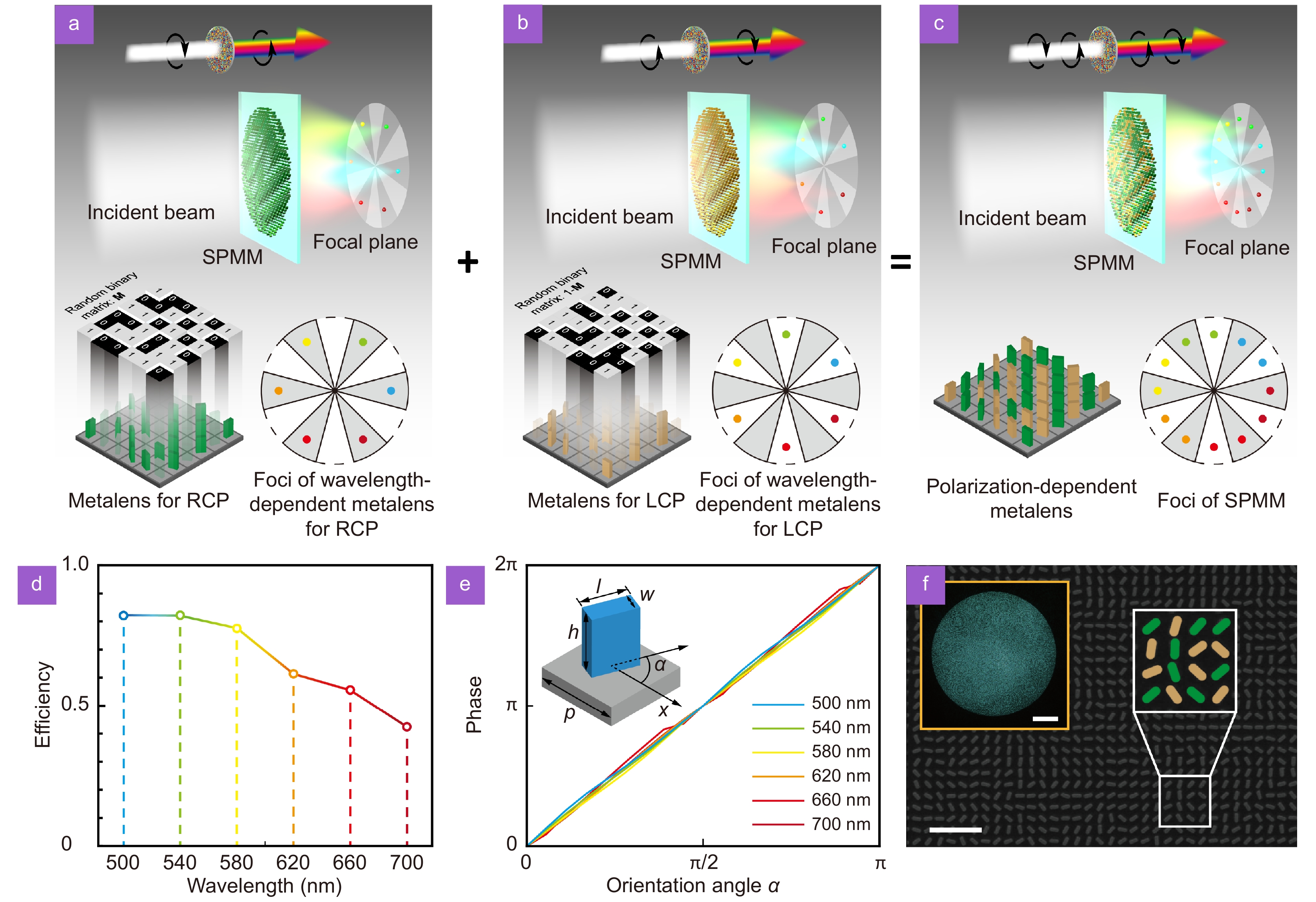
![Figure 2. Design and realization of the SPMM. (a, b) The metalenses designed for RCP (or LCP) based on the holography principle possess six working wavelengths and six corresponding off-axis foci at different positions on the same focal plane. (c) The SPMM with twelve different foci was acquired by mixing nanostructures of these two metalenses together randomly. (d, e) The simulated cross-polarization conversion efficiencies (d) and phase retardation (e) corresponding to six discrete wavelengths. The materials of the nanostructure and substrate were SiNx and SiO2, respectively. h=600 nm, w=130 nm, l=323 nm, and p=450 nm. (f) The SEM images of the fabricated metasurface (scale bar 2 μm). The fake color represents randomly mixed nanostructures. An optical inset shows the whole SPMM, which is 500 μm in diameter (scale bar 50 μm).]()
Design and realization of the SPMM. (a, b) The metalenses designed for RCP (or LCP) based on the holography principle possess six working wavelengths and six corresponding off-axis foci at different positions on the same focal plane. (c) The SPMM with twelve different foci was acquired by mixing nanostructures of these two metalenses together randomly. (d, e) The simulated cross-polarization conversion efficiencies (d) and phase retardation (e) corresponding to six discrete wavelengths. The materials of the nanostructure and substrate were SiNx and SiO2, respectively. h=600 nm, w=130 nm, l=323 nm, and p=450 nm. (f) The SEM images of the fabricated metasurface (scale bar 2 μm). The fake color represents randomly mixed nanostructures. An optical inset shows the whole SPMM, which is 500 μm in diameter (scale bar 50 μm).
In the design of SPMM, phase retardations induced by circular polarization conversion are achieved via space-variant anisotropic sub-wavelength structures. The same sub-wavelength structures generate the opposite phase for incident light beams with the opposite circular polarization state owing to the polarization-dependence of the geometric phase. Thus, a metalens with focal length f can be designed for any specific circular polarization and acquires a conjugate phase map of the lens under reverse-handedness circular polarized light incidence. The positive metalens designed for LCP light performs as negative metalens when illuminated by an RCP light beam, and vice versa. Indeed, the intensity of the focus on the focal plane resulting from the positive metalens is much higher than the divergent beam of the negative lens. To realize a polarization-dependent metalens with switchable foci, two metalenses with different working polarization states and off-axis foci are integrated. The sub-wavelength structures of positive metalenses for LCP or RCP light with corresponding off-axis foci are arranged in a rectangular distribution which can be treated as an abstractive “matrix”. Using this abstractive “RCP metalens matrix,” a random binary matrix M is generated to calculate the Hadamard product. In M, “0” and “1” are encountered with equal probability. Similarly, the result of the Hadamard product of the abstractive “LCP metalens matrix” and (1−M) can also be acquired. The polarization-dependent metalens design can be obtained by adding these two Hadamard product results together. The focal position of this metalens can be switched by changing the polarization of the incident light beam. In this design, we applied a random rather than a periodic binary matrix to avoid the potential grating diffraction effect. It should be noted that this randomly mixed metalens performs as a positive and a negative lens concurrently depending on whether the illuminating light beam is LCP or RCP. While, compared with the focus of the positive lens, the intensity of the divergent beam of the negative lens is too weak to observe at the focal plane and, therefore, can be ignored. Therefore, an SPMM with twelve foci can be obtained by combining two transversely dispersive metalenses randomly as a single metasurface element, as shown in Fig. 2(c).
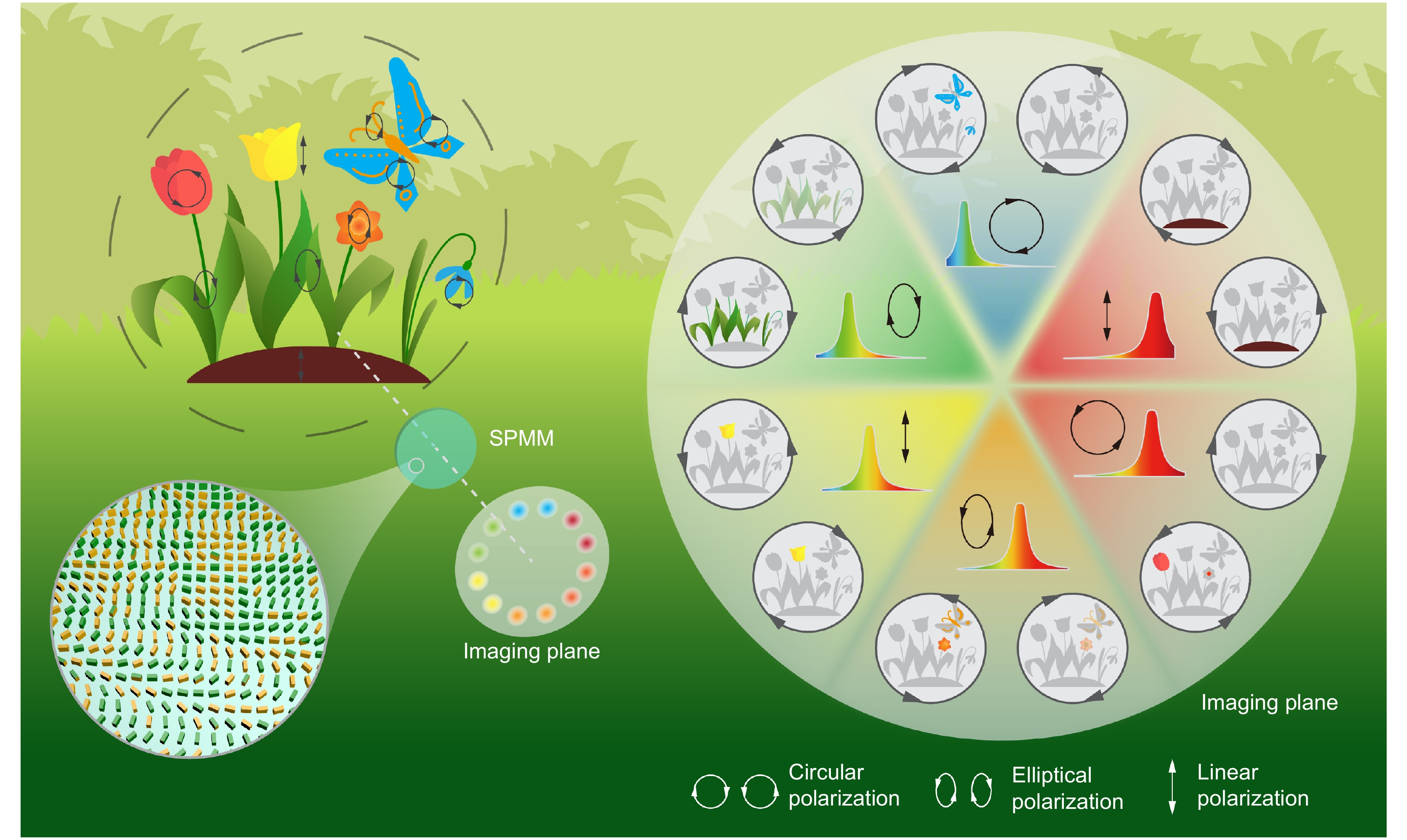
![Figure 1. Design concept of the SPMM device. The light from photographic scenes contains spectrum- and elliptic polarization-related information that is usually lost or ignored in traditional imaging systems. Our multi-foci metalens (inset) generates twelve foci/images corresponding to six spectral bands and two circular polarization states, thereby permitting the reconstruction of these lost features.]()
Design concept of the SPMM device. The light from photographic scenes contains spectrum- and elliptic polarization-related information that is usually lost or ignored in traditional imaging systems. Our multi-foci metalens (inset) generates twelve foci/images corresponding to six spectral bands and two circular polarization states, thereby permitting the reconstruction of these lost features.
Metalenses have been the subject of significant research interest in recent years35-39, in which considerable effort has been devoted to constructing single-foci metalenses. Moreover, due to the capability of complete light field control, it is possible to achieve multi-foci metalenses that can realize foci at different positions along the longitudinal or transverse axes using a single incident beam40-44. The design concept for our SPMM is illustrated in Fig. 1. The light from imaged objects contains rich information associated with multiple wavelengths and polarization ellipticity, which is usually lost or ignored in traditional intensity-based imaging methods. To address this issue, the SPMM generates twelve foci or images at different positions, which correspond to six bands of spectra and two orthogonal circular polarization states. Furthermore, the spectra and polarization ellipticity (linear, elliptical, or circular) relating to specific object areas can be resolved and reconstructed by identifying the focusing/imaging positions and corresponding relative intensities.
The SPMM presented herein consists of space-variant silicon nitride (SiNx) rectangular nanostructures. The phase retardation
where An and φn are the amplitude and phase of the nth positive lens that possesses the nth focus and nth working wavelength. The setting of amplitude factor An
for each profile is to adjust the weight of different wavelengths, e.g., to maintain the same conversion efficiencies for different wavelengths to construct high-performance devices, or to improve performance of metalens at a specific wavelength significantly. The focal lengths of each positive lens are identical, while the positions of each focus are different. In this paper, we demonstrate a metalens design with six working wavelengths and six corresponding off-axis foci at different positions. The foci positions of the left-handed circularly polarized (LCP) metalens are also different from those of the right-handed circularly polarized (RCP) metalens (Fig. 2(a, b)). SPMM-based focusing and imaging
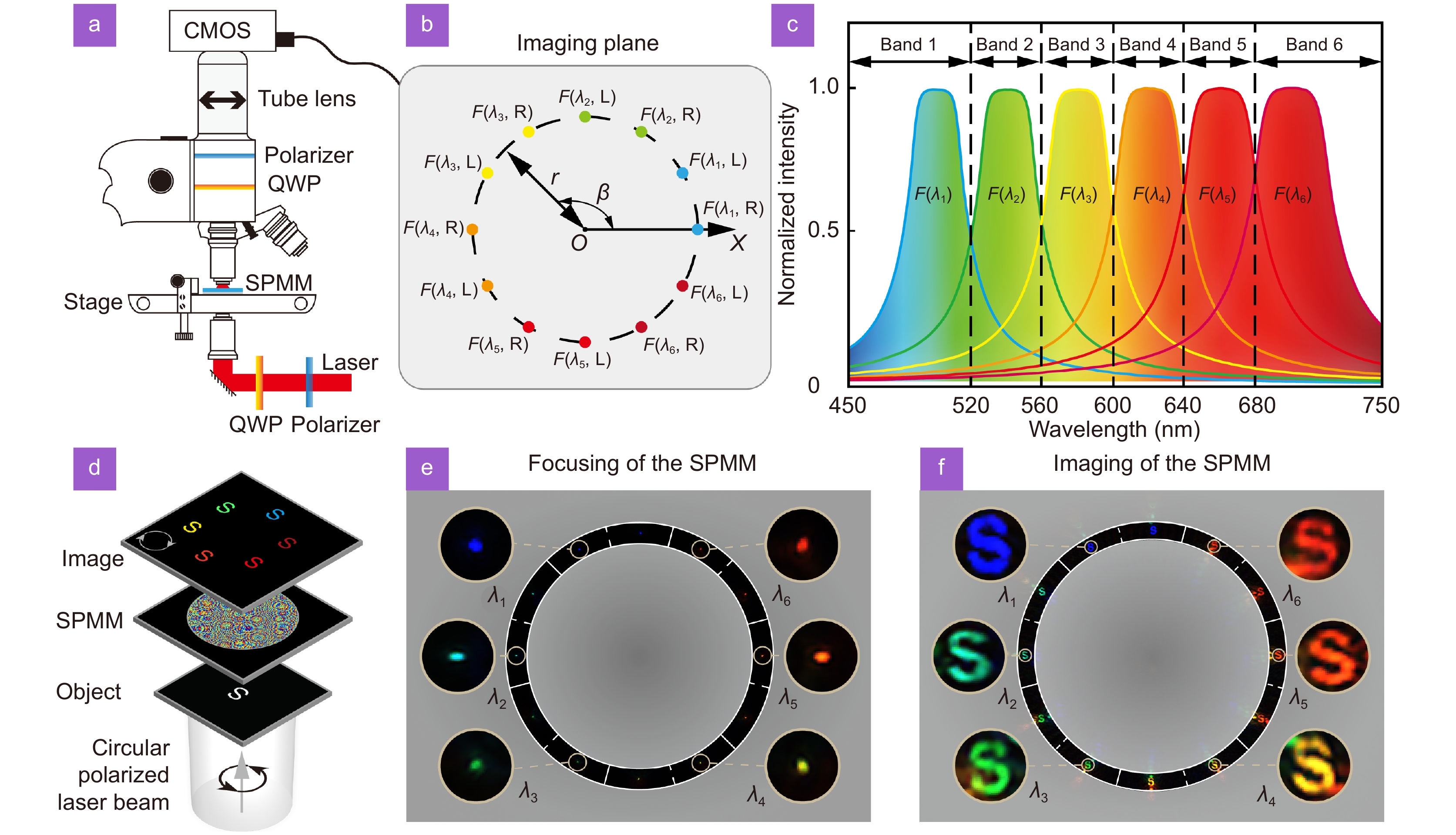
![Figure 3. Design and experimental focusing/imaging results for the SPMM. (a) The sketch map of optical setup with laser source (See Fig. S4(a) in Supplementary information for detailed description). (b) The positions of the twelve foci of the SPMM. (c) The wavelength dependency of the normalized total intensity of light spots at positions F(λn, L or R) within 10 μm. (d) Illustration of the experimental imaging progress. (e) The focusing results of the SPMM for the incident light beam consisted of six wavelengths. The diameter of each enlarged sliced illustration is 35 μm. (f) Typical imaging results of the SPMM for the incident light beam consisted of six wavelengths. The diameter of each sliced illustration is 35 μm.]()
Design and experimental focusing/imaging results for the SPMM. (a) The sketch map of optical setup with laser source (See Fig. S4(a) in Supplementary information for detailed description). (b) The positions of the twelve foci of the SPMM. (c) The wavelength dependency of the normalized total intensity of light spots at positions F(λn, L or R) within 10 μm. (d) Illustration of the experimental imaging progress. (e) The focusing results of the SPMM for the incident light beam consisted of six wavelengths. The diameter of each enlarged sliced illustration is 35 μm. (f) Typical imaging results of the SPMM for the incident light beam consisted of six wavelengths. The diameter of each sliced illustration is 35 μm.
As the SPMM design is based on six specific discrete wavelengths (λ1–λ6), the six light spots are all defocused when the wavelength of the incident beam is different from any of the designed wavelengths. However, the extent by which each spot is defocused differs, and there is still one light spot that is focused much better than the other five. For example, the total light intensity at F (λ1, L or R) is much higher than the other positions at the focal plane when the wavelength of the incident beam is approximately λ1. Therefore, the SPMM can also be used over a broad range to identify the spectral bands of the incident light beam (Fig. 3(c)). The focusing outcomes at six different positions on the focal plane (Fig. 3(e) and Fig. S5) demonstrate the multispectral focusing capability of the SPMM. Besides its focusing functionality, the SPMM can also achieve imaging functionality similar to that of a normal metalens. However, many differences remain between the imaging results acquired using the SPMM and a normal metalens. There are also six imaging positions I (λn, L or R) at the imaging plane, which are determined by the foci positions and object distances. The imaging process is illustrated in Fig. 3(d), with the typical experimental imaging results shown in Fig. 3(f) and Fig. S6. The results demonstrate SPMM’s distinguishing abilities of discrete wavelengths or spectral bands.
To show the foci and imaging results acquired by the SPMM for multi-wavelength incident light directly, a color CMOS camera was utilized to record experimental results at the focal or image plane. However, color CCD or CMOS cameras with spectral filters are unnecessary in practical applications because of the transversely dispersive space division property of the SPMM. The wavelengths of incident light beams can be distinguished by the positions rather than the colors of foci, even when using monochrome CCD or CMOS cameras. In fact, monochrome CCD/CMOS cameras are often more suitable for applications with higher requirements, such as astronomy and exploration, because they outperform color cameras in several aspects, such as higher quantum efficiency, lower read noise, and higher signal-to-noise ratio. Similarly, the classification of circular polarization states can be achieved by determining the positions of foci rather than using polarization analyzers. Thus, these two optical parameters (spectra and polarization ellipticity) can be distinguished simply by determining the positions of foci on the focal plane. Therefore, both theoretical and experimental results demonstrate the capability of the SPMM for resolving circular polarization states and finite discrete wavelengths (or spectral bands) with an ultra-compact design. Moreover, the widths and central wavelength distribution of each spectral band can be readily adjusted as required by changing the design parameters of the SPMM, such as the designed wavelength λn and the amplitude An.
by observing the position of the focus with the maximum light intensity at the focal plane, the polarization ellipticity and wavelength of the incident light beam can be determined. It should be noted that six focusing and imaging outcomes are obtained even with a single wavelength and one specific circular polarization because of the holography principle (see Section 2, Fig. S2, and Fig. S3 in Supplementary information for detailed information). However, there is only one light spot that is focused clearly on the focal plane, with the other five spots defocused owing to chromatic dispersion. In addition, the intensity of the focus at the position corresponding to the wavelength of incident light is much higher than that of the other five defocused light spots.
The positions of the twelve foci in our design are distributed evenly around a circle. The coordinates of the foci resulting from RCP and LCP incident light can be expressed as Eq. (2) and Eq. (3), respectively, using polar coordinates (Fig. 3(a, b)), where r=200 μm.
The measurement of the polarization ellipticity with SPMM
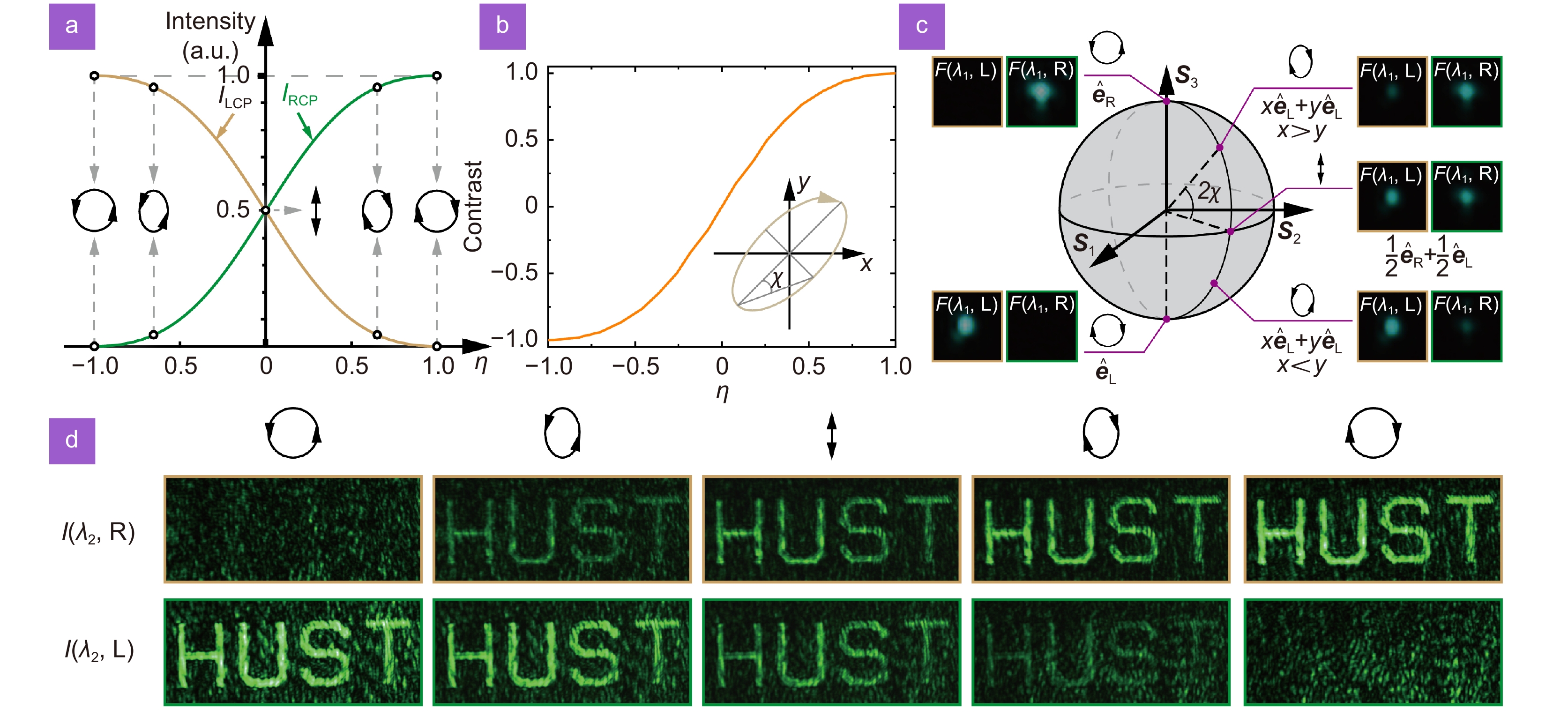
![Figure 4. Polarization-dependent focusing and imaging using the SPMM. (a) The relationship between normalized intensities IRCP (or ILCP) of RCP (or LCP) light and the polarization parameter η in the Jones matrices. (b) The contrast calculated by (IRCP−ILCP)/(IRCP+ILCP). (c) Typical polarization state measurement results at 500 nm, with the polarization state of the incident light beam varied gradually from LCP to RCP. The intensity of the focus at position F (500 nm, R) increases from zero to maximum, with this trend reversed at F (500 nm, L). (d) Imaging results achieved by the SPMM at a wavelength of 540 nm.]()
Polarization-dependent focusing and imaging using the SPMM. (a) The relationship between normalized intensities IRCP (or ILCP) of RCP (or LCP) light and the polarization parameter η in the Jones matrices. (b) The contrast calculated by (IRCP−ILCP)/(IRCP+ILCP). (c) Typical polarization state measurement results at 500 nm, with the polarization state of the incident light beam varied gradually from LCP to RCP. The intensity of the focus at position F (500 nm, R) increases from zero to maximum, with this trend reversed at F (500 nm, L). (d) Imaging results achieved by the SPMM at a wavelength of 540 nm.
and determined using the circular basis vectors
The SPMM can not only determine the rotational direction of circular polarization states but also characterize arbitrary polarization ellipticity (e.g., linear, elliptical, and circular) on a meridian of the Poincare sphere. The RCP and LCP states can be expressed as Eq. (4) and Eq. (5) using the respective Jones matrices.
where
Obviously, the experimental results in Fig. 3 correspond to the special conditions of
Assuming the longitude as constant, any polarization ellipticity on a meridian of the Poincare sphere can be described as
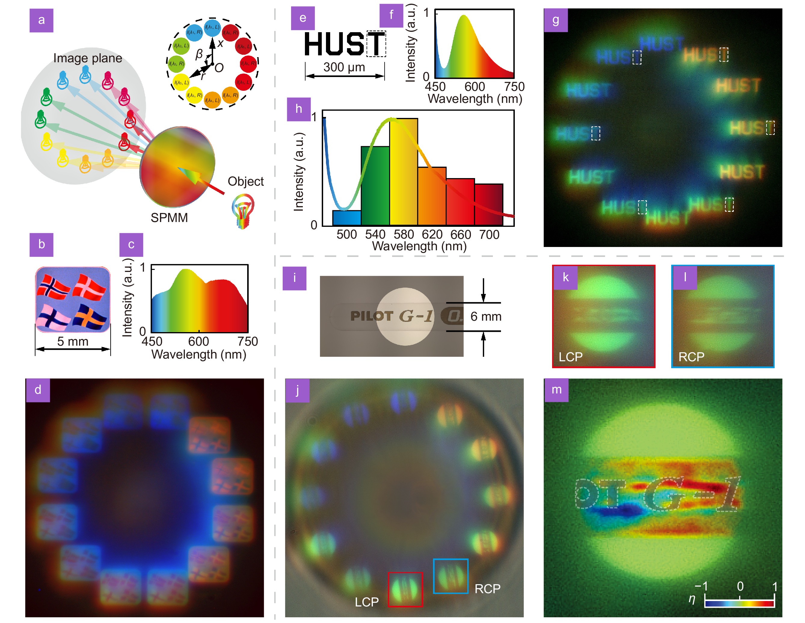
The polarization ellipticity resolved imaging of the SPMM also works well. The experimental imaging results at 540 nm are presented in Fig. 4(d). A "HUST" pattern left on an opaque coating substrate was used as the object to demonstrate the polarization ellipticity resolved imaging capability. Similar to the focusing process, the image positions are also polarization-dependent with a single wavelength because of the space division property of the SPMM at the imaging plane. The variation in the intensity of the two images at different positions (Fig. 4(d)) follows the same trend as observed for the focusing results in Fig. 4(c). The experimental results imply that the SPMM possesses the reconstruction capability of polarization ellipticity information in both focusing and imaging conditions.
The spectra and polarization ellipticity reconstruction of white light
To demonstrate the spectral reconstruction ability, the pattern of “HUST” was imaged using a normal flashlight from a mobile phone as the illumination source (Fig. 5(e–g)). The intensities of the typical part “T” marked with dashed lines in six images, were used to reconstruct the spectral histogram of the incident beam (Fig. 5(g)). It can be observed that most parts of the histogram fit well with the spectrum recorded by a spectrometer, which demonstrates the spectral reconstruction performance (Fig. 5(h)). However, the sixth band of the histogram is slightly higher because the cross-polarization conversion amplitude efficiency at λ6 = 700 nm is relatively low, leading to a low signal-to-noise ratio. This can be improved by optimizing the geometrical parameters and materials of the nanostructures.
where l1 and l2 represent object and image distances, respectively, and f is the focal length of the SPMM. Different flag colors in the same picture imply multiple spectra and imaging at different positions, thereby demonstrating the good multispectral imaging performance of the SPMM (see Fig. 5(d)).
![Figure 5. Multispectral and polarized imaging using the SPMM with ordinary white light beams. (a) Schematic illustration of the experiment. (b–d) Imaging results for a color picture consisting of four flags. The light source is a halogen lamp, and its spectrum is presented in (c). (e–g) Imaging results for the phrase]()
Multispectral and polarized imaging using the SPMM with ordinary white light beams. (a) Schematic illustration of the experiment. (b–d) Imaging results for a color picture consisting of four flags. The light source is a halogen lamp, and its spectrum is presented in (c). (e–g) Imaging results for the phrase "HUST", with the spectrum of a mobile phone flashlight that was used as the light source. (h) Histogram of the reconstructed spectra based on six regions marked with dash lines in (g). (i, j) Regular optical image and the SPMM imaging results for a transparent plastic stick. (k, l) A pair of typical images with the same spectral band are enlarged. (m) The reconstructed polarization ellipticity image can be calculated from the SPMM imaging results in (j). The images of corresponding opaque patterns are marked with dashed lines.
Although the experiments described in the previous section were performed using laser beams with discrete wavelengths, the SPMM can also work well with ordinary incoherent light with a wide spectrum from common light sources, such as the sun, a halogen lamp, or an LED (Fig. 5(a)). In this paper, we acquired images from several different ordinary light sources. A schematic of the experimental imaging system is shown in Fig. S4(b) of Supplementary information. Images of a colorful picture consisting of four flags (Fig. 5(b)) are shown in Fig. 5(d) under the illumination of the halogen light source with a wide spectrum (Fig. 5(c)). The size objects d is 5.0 mm. The object-image relationships can be calculated by
It can be seen that the SPMM can achieve spectra and polarization ellipticity reconstruction functions under ordinary light sources, which demonstrates its practice in general usages.
Furthermore, the polarization ellipticity image can be calculated using any pair of images corresponding to the same band of the spectrum (Fig. 5(i–m)). A transparent plastic stick taken from a common ballpoint pen was used to demonstrate our design (Fig. 5(i)). This transparent plastic stick exhibits complex stress birefringence, which cannot be captured by ordinary optical imaging. However, it can be observed through our SPMM by inspecting a pair of raw images in the same spectrum band (Fig. 5(j)). These two image copies (Fig. 5(k, l)) must be aligned, and the necessary distorted image rectification is applied to obtain the correct intensity distributions. The polarization ellipticity image can be obtained by the calculation represented in Eq. (8), and the fake color represents the polarization ellipticity factor
Conclusions
We have demonstrated the concept for SPMM capable of single-shot recognition and reconstruction of spectral and polarization ellipticity based on the geometric phase and holography principles, which has been verified experimentally with both coherent and incoherent light sources. As a transversely dispersive multi-foci metalens, SPMM generates 12 foci or images at different positions, which imply corresponding spectra and polarization ellipticity, thus providing a route toward the analysis of rich optical information by only a single shot. Compared with the existing special metasurface spectra- or polarization detection elements based on a micro-metalens array, the SPMM has a unique full-aperture design that can achieve superior focusing and imaging performance due to a significantly larger numerical aperture with the same fabricated size and focal length. In particular, through the demonstration of the SPMM imaging with both ordinary incoherent and coherent light sources, this work has exhibited its practical potential for the construction of ultra-compact multispectral and polarized imaging devices without the need of a multi-pass design using complicated spectral filters or mechanically moving parts. Moreover, this SPMM concept can be extended to the reconstruction of arbitrary points with both longitude and latitude on the Poincare sphere and achieve much finer partition of spectral bands via improved metalens design and nanofabrication techniques. Therefore, this work not only significantly reduces the size and weight of conventional optical devices but paves the way to achieve unprecedented recognition and reconstruction functionalities of optical information with an ultra-compact monolithic chip design. To be noted that the performance in some aspects of the multi-foci lens will become slightly worse than the usual single-focus lens with increased foci number. For example, the crosstalk issue of SPMM is a shortage needed to be optimized. However, the crosstalk issue can be mitigated in several ways, such as improving the arrangement of the imaging plane, setting specific imaging magnification, and utilizing various kinds of narrow-band spectral response nanostructures with different central wavelengths in the future. It is expected that such polarization ellipticity and wavelength-resolved SPMM could have a broad range of promising applications, including remote sensing, material characterization, optical communications, and information processing.
Methods
Fabrication of SPMM
The fabrication of the SPMM begins from a glass substrate with a thickness of 500 μm, as shown in Fig. S7 of Supplementary information. A silicon nitride thin film of 600 nm thickness is coated by plasma-enhanced chemical vapor deposition onto the substrate. Next, a chromium film of thickness 20 nm is deposited by electron-beam evaporation on the SiNx layer as a hard mask. Then a 200 nm photoresist layer (CSAR62) is spin-coated onto the top of the Cr layer. The nanostructure patterns are written by electron-beam lithography (Vistec: EBPG 5000 Plus) and implemented into the photoresist layer after development. The pattern is then transferred into the Cr hard mask layer by inductively coupled plasma etching (ICP, Oxford Plasmalab: System100-ICP-180), and the residual photoresist is stripped off by an oxygen plasma stripper (Diener electronic: PICO plasma stripper). Finally, the pattern is transferred into the SiNx layer by the next ICP process, and the remaining Cr is removed by Cr corrosion solution. The Cr layer is utilized as a hard mask because of the extremely high etching selectivity between Cr and SiNx.
Performance characterization of SPMM
The SPMM characterization with a laser source was performed using a home-built microscope (Fig. S4(a)). The light source used in the experiment is a supercontinuum broadband laser source (NKT SuperK, spectral range of 400–2000 nm) with a wavelength-selected modulator. The polarization states of the incident laser beam were modulated by changing the rotation angle of a linear polarizer and a quarter-wave plate. The transmitted light patterns were imaged by an objective lens and a tube lens, and recorded by a CMOS camera (Daheng Imaging, MER-U3). A cascaded quarter-wave plate and polarizer were used to filter the non-conversion background noise. For experiments with ordinary white light, the object is illuminated by a wide spectrum light source (halogen bulb of Motic microscope BA410 or camera flash light of Huawei Mate 20) (Fig. S4(b)).
Acknowledgements
We are grateful for financial supports from National Key Research and Development Program of China (Grant No. 2021YFF0502700), National Natural Science Foundation of China (Grant Nos. 52275429, 62205117, 61835008), Knowledge Innovation Program of Wuhan-Shuguang, Innovation project of Optics Valley Laboratory (Grant No. OVL2021ZD002), and Hubei Provincial Natural Science Foundation of China (Grant Nos. 2020CFA004, 2022CFB792).
Competing interests
The authors declare no competing financial interests.
-
References
[1] Tanré D, Bréon FM, Deuzé JL, Dubovik O, Ducos F et al. Remote sensing of aerosols by using polarized, directional and spectral measurements within the A-Train: the PARASOL mission. Atmos Meas Tech 4, 1383–1395 (2011).
DOI: 10.5194/amt-4-1383-2011[2] Yuen PWT, Richardson M. An introduction to hyperspectral imaging and its application for security, surveillance and target acquisition. Imaging Sci J 58, 241–253 (2010).
DOI: 10.1179/174313110X12771950995716[3] Liang HD. Advances in multispectral and hyperspectral imaging for archaeology and art conservation. Appl Phys A 106, 309–323 (2012).
DOI: 10.1007/s00339-011-6689-1[4] Levitt JA, Matthews DR, Ameer-Beg SM, Suhling K. Fluorescence lifetime and polarization-resolved imaging in cell biology. Curr Opin Biotechnol 20, 28–36 (2009).
DOI: 10.1016/j.copbio.2009.01.004[5] Park H, Crozier KB. Multispectral imaging with vertical silicon nanowires. Sci Rep 3, 2460 (2013).
DOI: 10.1038/srep02460[6] Zhao YQ, Yi C, Kong SG, Pan Q, Cheng YM. Multi-band Polarization Imaging and Applications (Springer, 2016).
Multi-band Polarization Imaging and Applications (Springer, 2016).
" target="_blank">Google ScholarView full references list -
Cited by
Periodical cited type(110)
1. Ahmed, H., Qi, B., Chen, X. Evolution of dispersion-engineered metasurfaces: Debye relaxation and folded path concept. Light Science and Applications, 2025, 14(1): 223. DOI:10.1038/s41377-025-01890-2 2. Yazdaanpanah, R., Ali, F., Korkmaz, S. et al. Polarization filtering with isomaterial multifocal metalenses at ultraviolet frequencies. Scientific Reports, 2025, 15(1): 22296. DOI:10.1038/s41598-025-04382-w 3. Ning, Y., Zhu, J., Ji, P. et al. Ultra-compact on-chip snapshot chiral spectral imaging with dispersion-engineered chiral metalens. 2025, 2(1): 23. DOI:10.1038/s44310-025-00066-5 4. Zhang, F., Bao, H., Pu, M. et al. Dispersion-engineered spin photonics based on folded-path metasurfaces. Light Science and Applications, 2025, 14(1): 198. DOI:10.1038/s41377-025-01850-w 5. Ai, Z., Yi, Y., Yang, H. et al. TiN-Ti ultra-broadband solar absorber: Efficient heat absorption and high thermal stability. Physics Letters Section A General Atomic and Solid State Physics, 2025. DOI:10.1016/j.physleta.2025.130827 6. Zhu, J., Liu, X., Li, A. et al. Opto-mechanical design of Fourier transform spectral polarization imaging system based on dual Wollaston prisms. Displays, 2025. DOI:10.1016/j.displa.2025.103026 7. Huang, Z., Wang, Z., Cheng, Y. et al. Metasurfaces empower optical multiparameter imaging: A review. Science China Physics Mechanics and Astronomy, 2025, 68(7): 274201. DOI:10.1007/s11433-025-2669-5 8. He, S., Tian, Y., Zhou, H. et al. Review for Micro-Nano Processing Technology of Microstructures and Metadevices. Advanced Functional Materials, 2025, 35(24): 2420369. DOI:10.1002/adfm.202420369 9. Deng, Z., Shi, Z., Huang, C. et al. Compact snapshot multispectral-depth imaging system with shared-modal multi-bandpass lenslet array (SMLA). Optics Letters, 2025, 50(11): 3568-3571. DOI:10.1364/OL.563215 10. Chen, F., Shi, S., Xue, S. et al. Recent Advances and Challenges in Multi-Photon 3D Nanoprinting. Engineering, 2025. DOI:10.1016/j.eng.2024.09.028 11. Dai, S., Bao, Y.-Q., Pan, J.-R. et al. A frequency-reconfigurable conformal metastructure absorber with liquid crystal temperature control for RCS reduction. Journal of Materials Chemistry C, 2025, 13(24): 12542-12553. DOI:10.1039/d5tc01304f 12. Li, D., Gu, M., Li, C. et al. The innovation in planar optics: Technological breakthroughs and application prospects of metalens. Precision Engineering, 2025. DOI:10.1016/j.precisioneng.2025.01.011 13. Liu, Y., Gu, M., Tian, Y. et al. 3D Printed Metamaterial Absorber Based on Vanadium Dioxide Phase Transition Control Prepared at Room Temperature. Laser and Photonics Reviews, 2025, 19(8): 2401673. DOI:10.1002/lpor.202401673 14. Chen, N., Zhou, X., Yin, L. et al. Angle-robust hyperspectral imaging based on quasi-random metal metasurfaces and cross-correlation optimization. Applied Optics, 2025, 64(11): 2949-2957. DOI:10.1364/AO.559959 15. Zheng, M.F., Tao, Y., Lou, B. et al. Additive Analysis of a Transmission-Type All-Dielectric Encoding Supergrating. JETP Letters, 2025, 121(7): 506-511. DOI:10.1134/S0021364025605494 16. Zhu, Z., Li, Y., Wang, J. et al. Reconfigurable origami chiral response for holographic imaging and information encryption. Opto Electronic Science, 2025, 4(4): 240026. DOI:10.29026/oes.2025.240026 17. Wu, P., Gu, M., Li, C. et al. Research progress on dynamic holographic display technology based on metasurfaces. Optics and Laser Technology, 2025. DOI:10.1016/j.optlastec.2024.112071 18. Xu, K., Liu, Y., Fan, X. et al. Simultaneous Dynamic Display of Meta-Hologram and Meta-Nanoprinting with High Frame Rate. Laser and Photonics Reviews, 2025, 19(6): 2400815. DOI:10.1002/lpor.202400815 19. Wang, L., Zhou, Y., Zhou, J. et al. Advances in On⁃Chip Infrared Polarization Imaging Devices Based on Micro⁃ and Nano⁃Structures (Invited) | [基 于 微 纳 结 构 的 片 上 红 外 偏 振 成 像 器 件 研 究 进 展(特 邀)]. Zhongguo Jiguang Chinese Journal of Lasers, 2025, 52(5): 0501002. DOI:10.3788/CJL241211 20. Zhou, J., Sun, X., Xu, J. et al. Versatile Tunable Terahertz Absorption Device Based on Bulk Dirac Semimetals and Graphene. Molecules, 2025, 30(5): 999. DOI:10.3390/molecules30050999 21. Jiang, J., Yi, Y., Sun, T. et al. A bi-directional metamaterial perfect absorber based on gold grating and TiO2-InAs normal hexagonal pattern film. Solar Energy Materials and Solar Cells, 2025. DOI:10.1016/j.solmat.2024.113330 22. Jia, M., Zhao, C., Wang, H. et al. Flexible terahertz beam manipulation and convolution operations in light-controllable digital coding metasurfaces. Iscience, 2025, 28(2): 111688. DOI:10.1016/j.isci.2024.111688 23. Zhao, F., Jing, X., Yu, M. Research progress on the principle and application of metalenses based on metasurfaces. Journal of Applied Physics, 2025, 137(5): 050701. DOI:10.1063/5.0246029 24. Meng, Z., Xiao, Y., Chen, L. et al. Floating Multi-Focus Metalens for High-Efficiency Airborne Laser Wireless Charging. Photonics, 2025, 12(2): 150. DOI:10.3390/photonics12020150 25. Shen, Z., Lv, B., Yi, Z. et al. Dual-frequency polarization-insensitive and wide-angle metasurface for electromagnetic energy harvesting. Photonics and Nanostructures Fundamentals and Applications, 2025. DOI:10.1016/j.photonics.2024.101328 26. Tang, Y., Zheng, S., Huang, W. et al. Polarization-Sensitive Deep-Groove Absorption Grating Based on Silicon. Physics of Wave Phenomena, 2025, 33(1): 46-56. DOI:10.3103/S1541308X24700523 27. He, Z., Fang, D., Yi, Y. Design of a Tunable Metamaterial Absorption Device with an Absorption Band Covering the Mid-Infrared Atmospheric Window. Photonics, 2025, 12(2): 148. DOI:10.3390/photonics12020148 28. Cen, C., Liu, X., Lin, Y. et al. Metamaterial absorber with ultra-broadband, ultra-high absorption, polarization independence and high-temperature resistance for solar thermal energy harvesting applications. Optics Communications, 2025. DOI:10.1016/j.optcom.2024.131292 29. Pu, M.B.. Integrated laser processing platform based on metasurface. Opto Electronic Advances, 2025, 8(2): 250017. DOI:10.29026/oea.2025.250017 30. Basilio-Ortiz, J.C., Moreno, I. Invariant optical properties of dielectric nanofins in geometric phase metasurfaces. Nanoscale, 2025. DOI:10.1039/d5nr02642c 31. Shang, Y., Qiao, S., Guo, Y. et al. Ultrasensitive Vector Displacement Measurement Based on Pancharatnam-Berry Phase Optical Element. Laser and Photonics Reviews, 2025. DOI:10.1002/lpor.202500741 32. Sun, T., Yang, Z., Cao, W. et al. Simultaneous 3D and Full Stokes Polarization Imaging Based on All-Dielectric Metalens Network. Laser and Photonics Reviews, 2025. DOI:10.1002/lpor.202500269 33. He, X., Lei, Y., Zhang, F. et al. Hybrid Meta-optics Enabled Ultra-Broadband and High-efficiency Polarization Imaging. Laser and Photonics Reviews, 2025. DOI:10.1002/lpor.202500623 34. Ma, Y., Liu, Y., He, Y. et al. Design of multipass cell with dense spot patterns and its performance in a light-induced thermoelastic spectroscopy-based methane sensor. Light Advanced Manufacturing, 2025, 6(1): 1. DOI:10.37188/lam.2025.001 35. Lu, K., Qi, J., Gu, M. et al. Microfluidic Biosensors Based on Dual-Layer Metasurfaces. IEEE Sensors Journal, 2025, 25(1): 419-425. DOI:10.1109/JSEN.2024.3496747 36. Huang, W.. Novel View Synthesis Based on Similar Perspective. Computer Animation and Virtual Worlds, 2025, 36(1): e70006. DOI:10.1002/cav.70006 37. Qiao, J., Li, C., Tian, Y. et al. Double-Validation Metasurface Holographic Encryption Based on XOR Algorithm. IEEE Transactions on Microwave Theory and Techniques, 2025, 73(3): 1741-1751. DOI:10.1109/TMTT.2024.3454062 38. Wang, W., Wang, L., Fu, Q. et al. Structural color: an emerging nanophotonic strategy for multicolor and functionalized applications. Opto Electronic Science, 2025, 4(4): 240030. DOI:10.29026/oes.2025.240030 39. Zhang, L., Ouyang, C., Wang, P. et al. Non-invasive in-situ monitoring of deep etching processes using terahertz metasurfaces. Optics Express, 2024, 32(26): 46999-47010. DOI:10.1364/OE.541662 40. Solati Masouleh, S., Hosseininejad, S.E. Terahertz beam shaping using space-time phase-only coded metasurfaces. Nano Communication Networks, 2024. DOI:10.1016/j.nancom.2024.100548 41. Ma, Y., Qiao, S., Wang, R. et al. A novel tapered quartz tuning fork-based laser spectroscopy sensing. Applied Physics Reviews, 2024, 11(4): 041412. DOI:10.1063/5.0214874 42. Li, Y., Liu, F., Zheng, M. et al. Reconfigurable metasurface design with hallucination and stealth capabilities. European Physical Journal D, 2024, 78(12): 148. DOI:10.1140/epjd/s10053-024-00938-z 43. Ke, L., Yang, Y., Zhu, M. et al. Tight focusing of fractional-order topological charge vector beams by cascading metamaterials and metalens. Microsystems and Nanoengineering, 2024, 10(1): 146. DOI:10.1038/s41378-024-00781-7 44. Qiao, J., Feng, G., Yao, G. et al. Research progress on the principle and application of multi-dimensional information encryption based on metasurface. Optics and Laser Technology, 2024. DOI:10.1016/j.optlastec.2024.111263 45. Wu, Y., Tao, W., Zhao, F. et al. Review for metamaterials and metasurfaces based on vanadium dioxide phase change materials. Optics and Laser Technology, 2024. DOI:10.1016/j.optlastec.2024.111376 46. Wang, X., Liu, W., Yi, Y. A three-band narrow-band terahertz perfect absorber for patch antennas and other sensors. Physics Letters Section A General Atomic and Solid State Physics, 2024. DOI:10.1016/j.physleta.2024.129887 47. Huang, W., Li, C., Tian, Y. Far field ring beam generation based on 3-bit encoded metasurface. Journal of Optics United Kingdom, 2024, 26(11): 115102. DOI:10.1088/2040-8986/ad820c 48. Cheng, X., Jin, Y., Tang, Y. et al. Image wireless transmission based on microwave digital coding metasurfaces. Journal of the Optical Society of America B Optical Physics, 2024, 41(11): D31-D39. DOI:10.1364/JOSAB.531277 49. Wang, X., Fan, X., Liu, Y. et al. 3D Nanolithography via Holographic Multi-Focus Metalens. Laser and Photonics Reviews, 2024, 18(11): 2400181. DOI:10.1002/lpor.202400181 50. He, X., Li, C., Fang, B. et al. Multichannel information transmission via a dual-frequency point space-time coding metasurface. Journal of the Optical Society of America B Optical Physics, 2024, 41(11): D15-D23. DOI:10.1364/JOSAB.530690 51. Tao, G., Zhao, Q., Song, Q. et al. A Polarization-Insensitive and Highly Sensitive THz Metamaterial Multi-Band Perfect Absorber. Micromachines, 2024, 15(11): 1388. DOI:10.3390/mi15111388 52. Chen, L., Yu, Y., Zhang, X. Imaging Spectropolarimeter Using a Multifunctional Metasurface. Nano Letters, 2024, 24(40): 12634-12641. DOI:10.1021/acs.nanolett.4c03787 53. Xu, N., Zhuge, Z., Li, H. et al. Color router-based long-wave infrared multispectral imaging. Optics Express, 2024, 32(21): 36875-36887. DOI:10.1364/OE.536948 54. Liu, Y., Tian, Y., Li, C. et al. All-dielectric double-layer honeycomb tunable metamaterial absorber with integrated gold nanoparticles. Photonics Research, 2024, 12(10): 2344-2353. DOI:10.1364/PRJ.533547 55. Jiang, S., Deng, W., Wang, Z. et al. Ka-Band metalens antenna empowered by physics-assisted particle swarm optimization (PA-PSO) algorithm. Opto Electronic Science, 2024, 3(10): 240014. DOI:10.29026/oes.2024.240014 56. Fu, J., Jiang, M., Wang, Z. et al. Supercritical metalens at h-line for high-resolution direct laser writing. Opto Electronic Science, 2024, 3(10): 230035. DOI:10.29026/oes.2024.230035 57. Liu, X., Li, Y., Yao, G. et al. Perfect vortex beams generation based on reflective geometric phase metasurfaces. Chinese Journal of Physics, 2024. DOI:10.1016/j.cjph.2024.08.028 58. Zhu, Z., Li, Y., Cui, F. et al. Coding Metasurface Beam Modulation Based on Phase Change Materials. Brazilian Journal of Physics, 2024, 54(5): 175. DOI:10.1007/s13538-024-01561-4 59. Luo, H., Zhu, Y., Song, Q. et al. Ultra-High-Efficiency Solar Capture Device Based on InAs Top Microstructure. Coatings, 2024, 14(10): 1297. DOI:10.3390/coatings14101297 60. Xin, H., Feng, G., Wang, Q. et al. Multi-channel vortex beam based on single layer transmission-type metal metasurface. Optics and Laser Technology, 2024. DOI:10.1016/j.optlastec.2024.111094 61. Tao, W., Wu, Y., Zhao, F. et al. Research progress in metamaterials and metasurfaces based on the phase change material Ge2Sb2Te5. Optics and Laser Technology, 2024. DOI:10.1016/j.optlastec.2024.111064 62. Gong, C., Yang, W., Cheng, S. et al. Design and performance study of a multiband metamaterial tunable thermal switching absorption device based on AlCuFe and VO2. Dalton Transactions, 2024, 53(40): 16647-16653. DOI:10.1039/d4dt01751j 63. Sun, H., Sun, T., Song, Q. et al. Temperature-tunable terahertz metamaterial device based on VO2 phase transition principle. Dalton Transactions, 2024, 53(42): 17299-17307. DOI:10.1039/d4dt02412e 64. Chen, B., Ma, C., Sun, T. et al. Three peak metamaterial broadband absorbing materials based on ZnSe-Cr-InAs stacked disk arrays. Dalton Transactions, 2024, 53(42): 17291-17298. DOI:10.1039/d4dt01971g 65. Xu, X., Tang, F., Zhang, X. et al. Unveiling the Terahertz Nano-Fingerprint Spectrum of Single Artificial Metallic Resonator. Sensors, 2024, 24(18): 5866. DOI:10.3390/s24185866 66. Dai, T., Yi, Y., Yi, Z. et al. Photonic Crystal Fiber Based on Surface Plasmon Resonance Used for Two Parameter Sensing for Magnetic Field and Temperature. Photonics, 2024, 11(9): 784. DOI:10.3390/photonics11090784 67. Liu, W., Wu, F., Yi, Z. et al. Broadband Solar Absorber and Thermal Emitter Based on Single-Layer Molybdenum Disulfide. Molecules, 2024, 29(18): 4515. DOI:10.3390/molecules29184515 68. Xu, T., Zhang, W., Song, Q. et al. Thermotunable mid-infrared metamaterial absorption material based on combined hollow cylindrical VO2 structure. Surfaces and Interfaces, 2024. DOI:10.1016/j.surfin.2024.104868 69. Zheng, R., Yi, Y., Song, Q. et al. Dual-Tuned Terahertz Absorption Device Based on Vanadium Dioxide Phase Transition Properties. Materials, 2024, 17(17): 4287. DOI:10.3390/ma17174287 70. Wu, Z., Teng, Z., Xu, K. et al. Meta-Holography: Dynamic Display, Encryption, Imaging, and Others | [超 表 面 全 息 术:动 态 显 示 、加 密 、成 像 及 其 他]. Zhongguo Jiguang Chinese Journal of Lasers, 2024, 51(17): 1700003. DOI:10.3788/CJL240843 71. Zhu, J., Li, C., Fang, B. et al. Advancements in biosensing detection based on terahertz metasurfaces. Optics and Laser Technology, 2024. DOI:10.1016/j.optlastec.2024.110978 72. Guo, K., Yang, D., Liu, B. et al. Random broadband filters based on combination of metasurface and multilayer thin films for hyperspectral imaging. Journal of Physics D Applied Physics, 2024, 57(31): 315103. DOI:10.1088/1361-6463/ad4370 73. Li, A., Wu, Y., Wang, C. et al. An inversely designed integrated spectrometer with reconfigurable performance and ultra-low power consumption. Opto Electronic Advances, 2024, 7(8): 240099. DOI:10.29026/oea.2024.240099 74. Yang, Z., Wang, X., Jin, Y. et al. Calculation and experiment of carpet illusionary cloaking modeling based on coding metasurface. Sensors and Actuators A Physical, 2024. DOI:10.1016/j.sna.2024.115395 75. Liu, L., Liu, W., Fu, W. et al. Enhancing the Overall Performance of Perovskite Solar Cells with a Nano-Pyramid Anti-Reflective Layer. Photonics, 2024, 11(8): 715. DOI:10.3390/photonics11080715 76. Xu, J., Xu, P., Yang, Z. et al. Freeform metasurface design with a conditional generative adversarial network. Applied Physics A Materials Science and Processing, 2024, 130(8): 530. DOI:10.1007/s00339-024-07694-2 77. Chen, Y., Zhang, S., Tian, Y. et al. Focus control of wide-angle metalens based on digitally encoded metasurface. Opto Electronic Advances, 2024, 7(8): 240095. DOI:10.29026/oea.2024.240095 78. Zheng, S., Yu, Y., Zhou, X. et al. Research on Multidimensional Optical Imaging Empowered by Metasurfaces (Invited) | [超 构 表 面 赋 能 多 维 度 光 学 成 像 研 究(特 邀)]. Laser and Optoelectronics Progress, 2024, 61(16): 1611001. DOI:10.3788/LOP241403 79. Liu, Y., Zhang, T., Cai, J. et al. Simultaneous Circular Dichroism and Wavefront Manipulation with Generalized Pancharatnam-Berry Phase Metasurfaces. ACS Applied Materials and Interfaces, 2024, 16(26): 33935-33942. DOI:10.1021/acsami.4c03439 80. Hou, J., Liu, X., Liu, Y. et al. Highly sensitive CO2-LITES sensor based on a self-designed low-frequency quartz tuning fork and fiber-coupled MPC. Chinese Optics Letters, 2024, 22(7): 073001. DOI:10.3788/COL202422.073001 81. Li, Y., Xu, J., Liu, F. et al. Design and Analysis of Terahertz Wave Tunable Metamaterial with High Refractive Index. Silicon, 2024, 16(11): 4621-4633. DOI:10.1007/s12633-024-03022-8 82. Chen, P., Yi, Y., Song, Q. et al. Simulation and Analysis of a Near-Perfect Solar Absorber Based on SiO2-Ti Cascade Optical Cavity. Photonics, 2024, 11(7): 604. DOI:10.3390/photonics11070604 83. Xu, T., Yi, Y., Song, Q. et al. Design of a Far-Infrared Broadband Metamaterial Absorber with High Absorption and Ultra-Broadband. Coatings, 2024, 14(7): 799. DOI:10.3390/coatings14070799 84. Li, Y., Xu, J., Liu, F. et al. Broadband achromatic transmission stealth cloak based on all dielectric metasurfaces. Physica Scripta, 2024, 99(7): 075536. DOI:10.1088/1402-4896/ad5803 85. Cui, F., Huang, X., Zhou, Q. et al. Magnetic toroidal dipole resonance terahertz wave biosensor based on all-silicon metasurface. Optics and Lasers in Engineering, 2024. DOI:10.1016/j.optlaseng.2024.108128 86. Yang, Y., Xin, H., Liu, Y. et al. Intelligent metasurfaces: Integration of artificial intelligence technology and metasurfaces. Chinese Journal of Physics, 2024. DOI:10.1016/j.cjph.2024.03.043 87. Zhou, P., Zhou, J., Lv, Y. Metasurface CGH optimization for CTIS based on interior point method. Laser Physics Letters, 2024, 21(5): 055207. DOI:10.1088/1612-202X/ad3815 88. Wang, S., Zeng, D., Xu, Y. et al. Towards complex scenes: A deep learning-based camouflaged people detection method for snapshot multispectral images. Defence Technology, 2024. DOI:10.1016/j.dt.2023.12.011 89. Cheng, X., Li, C., Fang, B. et al. Metasurface-based wireless communication technology and its applications. Journal of Applied Physics, 2024, 135(12): 120702. DOI:10.1063/5.0198211 90. Deng, Y., Huang, X., Lu, Z. et al. Multifunctional metalens optical tweezers for optical information recognition. Optics Express, 2024, 32(6): 9456-9467. DOI:10.1364/OE.516792 91. Lou, B., Xu, J., Liu, F. et al. Scattering control based on geometric phase reflection-type coded metasurface. Optoelectronics and Advanced Materials Rapid Communications, 2024, 18(3-4): 113-119. 
92. Zhao, Y., Liu, F., Sui, Z. et al. Circular-target-style bifocal zoom metalens. Optics Express, 2024, 82(3): 3241-3250. DOI:10.1364/OE.514548 93. Fan, Q., Xu, W., Lin, P. et al. Aluminum-based plasmonic metasurface for computational spectrometry with full coverage of visible light. Optics Letters, 2024, 49(1): 21-24. DOI:10.1364/OL.503626 94. Ma, M., Chen, R., Chen, C. et al. Silicon Based Integrated Plasmonic Schottky Spectral-3olarization Detector Array. 2024. DOI:10.1109/3M-NANO61605.2024.10769684 95. Yang, Y., Jiang, J., Li, C. et al. Flexible Control of Encoded Metasurface Holographic Imaging Based on Fourier Convolution Principle. Journal of Lightwave Technology, 2024, 42(21): 7516-7522. DOI:10.1109/JLT.2024.3414153 96. Zhang, X., Chen, Q., Tang, D. et al. Broadband high-efficiency dielectric metalenses based on quasi-continuous nanostrips. Opto Electronic Advances, 2024, 7(5): 230126. DOI:10.29026/oea.2024.230126 97. Zhang, J.C., Chen, M.K., Fan, Y. et al. Miniature tunable Airy beam optical meta-device. Opto Electronic Advances, 2024, 7(2): 230171. DOI:10.29026/oea.2024.230171 98. Sun, H., He, Y., Qiao, S. et al. Highly sensitive and real-simultaneous CH4/C2H2 dual-gas LITES sensor based on Lissajous pattern multi-pass cell. Opto Electronic Science, 2024, 3(11): 240013. DOI:10.29026/oes.2024.240013 99. Liu, Y., Li, C., Tang, Y. et al. Terahertz Wave all-Dielectric Broadband Tunable Metamaterial Absorber. Journal of Lightwave Technology, 2024, 42(21): 7686-7692. DOI:10.1109/JLT.2024.3376349 100. Chen, Y., Huang, X., Yao, G. et al. Microfluidic Sensor Based on Substrate-Free Non-Uniform Metagrating. Journal of Lightwave Technology, 2024, 42(21): 7498-7506. DOI:10.1109/JLT.2024.3405198 101. Gao, H., Fan, X., Wang, Y. et al. Metasurface-Based Orbital Angular Momentum Multi-Dimensional Demultiplexer and Decoder. Laser and Photonics Reviews, 2023, 17(12): 2300393. DOI:10.1002/lpor.202300393 102. Hou, E., Liang, Z., Shi, X. et al. All-dielectric six-foci metalens for infrared polarization detection based on Stokes space. Optics Express, 2023, 31(24): 40018-40028. DOI:10.1364/OE.504936 103. Xiao, Y., Chen, L., Pu, M. et al. Improved spatiotemporal resolution of anti-scattering super-resolution label-free microscopy via synthetic wave 3D metalens imaging. Opto Electronic Science, 2023, 2(11) DOI:10.29026/oes.2023.230037 104. Wang, X., Xu, K., Fan, X. et al. Transversely Dispersive Multi-Foci Metalens Based on Selective Spectral Response Structure | [基 于 选 择 性 光 谱 响 应 结 构 的 横 向 色 散 多 焦 点 超 透 镜]. Zhongguo Jiguang Chinese Journal of Lasers, 2023, 50(18): 1813014. DOI:10.3788/CJL230941 105. Atalay Appak, I.A., Sahin, E., Guillemot, C. et al. Learning flat optics for extended depth of field microscopy imaging. Nanophotonics, 2023, 12(18): 3623-3632. DOI:10.1515/nanoph-2023-0321 106. Shen, S., Li, S., Yuan, Y. et al. High-efficiency broadband achromatic metalenses for visible full-stokes polarization imaging. Optics Express, 2023, 31(17): 28611-28623. DOI:10.1364/OE.496492 107. Li, Y., Huang, X., Liu, S. et al. Metasurfaces for near-eye display applications. Opto Electronic Science, 2023, 2(8): 230025. DOI:10.29026/oes.2023.230025 108. Ali, J., Ahmad, A., Choi, D.-Y. A Metalens Design for On- and Off-Center Focusing with Amorphous Silicon Hydrogenated (a-Si:H)-Based 1D Array in Visible Spectrum. Electronics Switzerland, 2023, 12(13): 2953. DOI:10.3390/electronics12132953 109. Rui, F., Yaokai, T., Yalong, L. et al. Polarization-multiplexed optical differentiation using topological metasurfaces | [拓扑优化超表面的偏振复用光学微分运算]. Guangdian Gongcheng Opto Electronic Engineering, 2023, 50(9): 230172. DOI:10.12086/oee.2023.230172 110. Wan, Y., Liu, W., Lin, R. et al. Research progress and applications of spectral imaging based on metasurfaces | [基于超构表面的光谱成像及应用研究进展]. Guangdian Gongcheng Opto Electronic Engineering, 2023, 50(8): 230139. DOI:10.12086/oee.2023.230139 Other cited types(0)
-
Author Information
-
Supplementary Information
TitleActions -
Copyright
Open Access. © The Author(s). This article is licensed under a Creative Commons Attribution 4.0 International License. To view a copy of this license, visit http://creativecommons.org/licenses/by/4.0/. -
About this Article
Cite this Article
Hui Gao, Xuhao Fan, Yuxi Wang, et al. Multi-foci metalens for spectra and polarization ellipticity recognition and reconstruction. Opto-Electronic Science 2, 220026 (2023). DOI: 10.29026/oes.2023.220026Download CitationArticle History
- Received Date December 04, 2022
- Revised Date January 04, 2023
- Accepted Date January 12, 2023
- Published Date March 29, 2023
Article Metrics
Article Views(9878) PDF Downloads(1071)
New website getting online, testing
 OE Journals Group

Reports the physical mechanisms and fundamental science of optics, photonics and optoelectronics
Open Access