| Citation: | Jing JY, Liu K, Jiang JF, Xu TH, Wang S et al. Highly sensitive and stable probe refractometer based on configurable plasmonic resonance with nano-modified fiber core. Opto-Electron Adv 6, 220072 (2023). doi: 10.29026/oea.2023.220072 |
Highly sensitive and stable probe refractometer based on configurable plasmonic resonance with nano-modified fiber core
-
Abstract
A dispersion model is developed to provide a generic tool for configuring plasmonic resonance spectral characteristics. The customized design of the resonance curve aiming at specific detection requirements can be achieved. According to the model, a probe-type nano-modified fiber optic configurable plasmonic resonance (NMF-CPR) sensor with tip hot spot enhancement is demonstrated for the measurement of the refractive index in the range of 1.3332–1.3432 corresponding to the low-concentration biomarker solution. The new-type sensing structure avoids excessive broadening and redshift of the resonance dip, which provides more possibilities for the surface modification of other functional nanomaterials. The tip hot spots in nanogaps between the Au layer and Au nanostars (AuNSs), the tip electric field enhancement of AuNSs, and the high carrier mobility of the WSe2 layer synergistically and significantly enhance the sensitivity of the sensor. Experimental results show that the sensitivity and the figure of merit of the tip hot spot enhanced fiber NMF-CPR sensor can achieve up to 2995.70 nm/RIU and 25.04 RIU−1, respectively, which are 1.68 times and 1.29 times higher than those of the conventional fiber plasmonic resonance sensor. The results achieve good agreements with numerical simulations, demonstrate a better level compared to similar reported studies, and verify the correctness of the dispersion model. The detection resolution of the sensor reaches up to 2.00×10⁻⁵ RIU, which is obviously higher than that of the conventional side-polished fiber plasmonic resonance sensor. This indicates a high detection accuracy of the sensor. The dense Au layer effectively prevents the intermediate nanomaterials from shedding and chemical degradation, which enables the sensor with high stability. Furthermore, the terminal reflective sensing structure can be used as a practical probe and can allow a more convenient operation. -

-
References
[1] Liu T, Li H, He T, Fan CZ, Yan ZJ et al. Ultra-high resolution strain sensor network assisted with an LS-SVM based hysteresis model. Opto-Electron Adv 4, 200037 (2021). doi: 10.29026/oea.2021.200037 [2] Guan BO, Jin L, Ma J, Liang YZ, Bai X. Flexible fiber-laser ultrasound sensor for multiscale photoacoustic imaging. Opto-Electron Adv 4, 200081 (2021). doi: 10.29026/oea.2021.200081 [3] Wang RL, Zhang HZ, Liu QY, Liu F, Han XL et al. Operando monitoring of ion activities in aqueous batteries with plasmonic fiber-optic sensors. Nat Commun 13, 547 (2022). doi: 10.1038/s41467-022-28267-y [4] Caucheteur C, Guo T, Liu F, Guan BO, Albert J. Ultrasensitive plasmonic sensing in air using optical fibre spectral combs. Nat Commun 7, 13371 (2016). doi: 10.1038/ncomms13371 [5] Lee C, Lawrie B, Pooser R, Lee KG, Rockstuhl C et al. Quantum plasmonic sensors. Chem Rev 121, 4743–4804 (2021). doi: 10.1021/acs.chemrev.0c01028 [6] Liu Y, Peng W. Fiber-optic surface Plasmon resonance sensors and biochemical applications: a review. J Lightwave Technol 39, 3781–3791 (2021). doi: 10.1109/JLT.2020.3045068 [7] Zhao Y, Tong RJ, Xia F, Peng Y. Current status of optical fiber biosensor based on surface Plasmon resonance. Biosens Bioelectron 142, 111505 (2019). doi: 10.1016/j.bios.2019.111505 [8] Jing JY, Liu K, Jiang JF, Xu TH, Wang S et al. Performance improvement approaches for optical fiber SPR sensors and their sensing applications. Photonics Res 10, 126–147 (2022). doi: 10.1364/PRJ.439861 [9] Singh Y, Raghuwanshi SK. Titanium dioxide (TiO2) coated optical fiber-based SPR sensor in near-infrared region with bimetallic structure for enhanced sensitivity. Optik 226, 165842 (2021). doi: 10.1016/j.ijleo.2020.165842 [10] Li LK, Zhang YN, Zhou YF, Zheng WL, Sun YT et al. Optical fiber optofluidic bio-chemical sensors: a review. Laser Photonics Rev 15, 2000526 (2021). doi: 10.1002/lpor.202000526 [11] Singh R, Kumar S, Liu FZ, Shuang C, Zhang BY et al. Etched multicore fiber sensor using copper oxide and gold nanoparticles decorated graphene oxide structure for cancer cells detection. Biosens Bioelectron 168, 112557 (2020). doi: 10.1016/j.bios.2020.112557 [12] Wang GY, Lu Y, Duan LC, Yao JQ. A refractive index sensor based on PCF with ultra-wide detection range. IEEE J Sel Top Quant 27, 5600108 (2021). doi: 10.1109/JSTQE.2020.2993866 [13] Urrutia A, Del Villar I, Zubiate P, Zamarreño CR. A comprehensive review of optical fiber refractometers: toward a standard comparative criterion. Laser Photonics Rev 13, 1900094 (2019). doi: 10.1002/lpor.201900094 [14] Liu LH, Zhang XJ, Zhu Q, Li KW, Lu Y et al. Ultrasensitive detection of endocrine disruptors via superfine plasmonic spectral combs. Light Sci Appl 10, 181 (2021). doi: 10.1038/s41377-021-00618-2 [15] Weng SJ, Pei L, Wang JS, Ning TG, Li J. High sensitivity D-shaped hole fiber temperature sensor based on surface Plasmon resonance with liquid filling. Photonics Res 5, 103–107 (2017). doi: 10.1364/PRJ.5.000103 [16] Wang D, Li W, Zhang QR, Liang BQ, Peng ZK et al. High-performance tapered fiber surface Plasmon resonance sensor based on the graphene/Ag/TiO2 layer. Plasmonics 16, 2291–2303 (2021). doi: 10.1007/s11468-021-01483-w [17] Song H, Wang Q, Zhao WM. A novel SPR sensor sensitivity-enhancing method for immunoassay by inserting MoS2 nanosheets between metal film and fiber. Opt Laser Eng 132, 106135 (2020). doi: 10.1016/j.optlaseng.2020.106135 [18] Jing JY, Liu K, Jiang JF, Xu TH, Wang S et al. Double-antibody sandwich immunoassay and plasmonic coupling synergistically improved long-range SPR biosensor with low detection limit. Nanomaterials 11, 2137 (2021). doi: 10.3390/nano11082137 [19] Chien FC, Chen SJ. A sensitivity comparison of optical biosensors based on four different surface Plasmon resonance modes. Biosens Bioelectron 20, 633–642 (2004). doi: 10.1016/j.bios.2004.03.014 [20] Jain S, Paliwal A, Gupta V, Tomar M. Smartphone integrated handheld Long Range Surface Plasmon Resonance based fiber-optic biosensor with tunable SiO2 sensing matrix. Biosens Bioelectron 201, 113919 (2022). doi: 10.1016/j.bios.2021.113919 [21] Shrivastav AM, Satish L, Kushmaro A, Shvalya V, Cvelbar U et al. Engineering the penetration depth of nearly guided wave surface Plasmon resonance towards application in bacterial cells monitoring. Sensor Actuators B:Chem 345, 130338 (2021). doi: 10.1016/j.snb.2021.130338 [22] Jiang XD, Xu WR, Ilyas N, Li MC, Guo RK et al. High-Performance coupled Plasmon waveguide resonance optical sensor based on SiO2: Ag film. Results Phys 26, 104308 (2021). doi: 10.1016/j.rinp.2021.104308 [23] Ma JY, Liu K, Jiang JF, Xu TH, Wang S et al. All optic-fiber coupled Plasmon waveguide resonance sensor using ZrS2 based dielectric layer. Opt Express 28, 11280–11289 (2020). doi: 10.1364/OE.389279 [24] Ji LT, Yang SQ, Shi RN, Fu YJ, Su J et al. Polymer waveguide coupled surface Plasmon refractive index sensor: a theoretical study. Photonic Sens 10, 353–363 (2020). doi: 10.1007/s13320-020-0589-y [25] Ross MB, Ku JC, Lee B, Mirkin CA, Schatz GC. Plasmonic metallurgy enabled by DNA. Adv Mater 28, 2790–2794 (2016). doi: 10.1002/adma.201505806 [26] Zhang WJ, Zeng XL, Yang A, Teng LP, Zhu Y. Research on evanescent field ammonia detection with gold-nanosphere coated microfibers. Opto-Electron Eng 48, 200451 (2021). [27] Jing JY, Zhu Q, Dai ZX, Li SY, Wang Q et al. Sensing self-referenced fiber optic long-range surface Plasmon resonance sensor based on electronic coupling between surface Plasmon polaritons. Appl Opt 58, 6329–6334 (2019). doi: 10.1364/AO.58.006329 [28] Yang YL, Chen HJ, Zou XX, Shi XL, Liu WD et al. Flexible carbon-fiber/semimetal Bi nanosheet arrays as separable and recyclable plasmonic photocatalysts and photoelectrocatalysts. ACS Appl Mater Interfaces 12, 24845–24854 (2020). doi: 10.1021/acsami.0c05695 [29] Kabashin AV, Evans P, Pastkovsky S, Hendren W, Wurtz GA et al. Plasmonic nanorod metamaterials for biosensing. Nat Mater 8, 867–871 (2009). doi: 10.1038/nmat2546 [30] Kant R, Tabassum R, Gupta BD. Xanthine oxidase functionalized Ta2O5 nanostructures as a novel scaffold for highly sensitive SPR based fiber optic xanthine sensor. Biosens Bioelectron 99, 637–645 (2018). doi: 10.1016/j.bios.2017.08.040 [31] Chen JN, Badioli M, Alonso-González P, Thongrattanasiri S, Huth F et al. Optical nano-imaging of gate-tunable graphene plasmons. Nature 487, 77–81 (2012). doi: 10.1038/nature11254 [32] Singh Y, Paswan MK, Raghuwanshi SK. Sensitivity enhancement of SPR sensor with the black phosphorus and graphene with Bi-layer of gold for chemical sensing. Plasmonics 16, 1781–1790 (2021). doi: 10.1007/s11468-020-01315-3 [33] Liu LX, Ye K, Jia ZY, Xue TY, Nie AM et al. High-sensitivity and versatile plasmonic biosensor based on grain boundaries in polycrystalline 1L WS2 films. Biosens Bioelectron 194, 113596 (2021). doi: 10.1016/j.bios.2021.113596 [34] Wu Q, Li NB, Wang Y, Xu YC, Wu JD et al. Ultrasensitive and selective determination of carcinoembryonic antigen using multifunctional ultrathin amino-functionalized Ti3C2-MXene nanosheets. Anal Chem 92, 3354–3360 (2020). doi: 10.1021/acs.analchem.9b05372 [35] Yang S, Bao W, Liu XZ, Kim J, Zhao RK et al. Subwavelength-scale lasing perovskite with ultrahigh Purcell enhancement. Matter 4, 4042–4050 (2021). doi: 10.1016/j.matt.2021.10.024 [36] Shalabney A, Abdulhalim I. Sensitivity-enhancement methods for surface Plasmon sensors. Laser Photonics Rev 5, 571–606 (2011). doi: 10.1002/lpor.201000009 [37] Zhao Y, Lei M, Liu SX, Zhao Q. Smart hydrogel-based optical fiber SPR sensor for pH measurements. Sensor Actuators B:Chem 261, 226–232 (2018). doi: 10.1016/j.snb.2018.01.120 [38] Zhao ZH, Wang Q. Gold nanoparticles (AuNPs) and graphene oxide heterostructures with gold film coupling for an enhanced sensitivity surface Plasmon resonance (SPR) fiber sensor. Instrum Sci Technol 50, 530–542 (2022). doi: 10.1080/10739149.2022.2044845 [39] Kravets VG, Kabashin AV, Barnes WL, Grigorenko AN. Plasmonic surface lattice resonances: a review of properties and applications. Chem Rev 118, 5912–5951 (2018). doi: 10.1021/acs.chemrev.8b00243 [40] Shi S, Wang LB, Su RX, Liu BS, Huang RL et al. A polydopamine-modified optical fiber SPR biosensor using electroless-plated gold films for immunoassays. Biosens Bioelectron 74, 454–460 (2015). doi: 10.1016/j.bios.2015.06.080 [41] Chiavaioli F, Gouveia CAJ, Jorge PAS, Baldini F. Towards a uniform metrological assessment of grating-based optical fiber sensors: from refractometers to biosensors. Biosensors 7, 23 (2017). doi: 10.3390/bios7020023 [42] Zhang XZ, Yang BY, Jiang JF, Liu K, Fan XJ et al. Side-polished SMS based RI sensor employing macro-bending perfluorinated POF. Opto-Electron Adv 4, 200041 (2021). doi: 10.29026/oea.2021.200041 [43] Dastmalchi B, Tassin P, Koschny T, Soukoulis CM. A new perspective on plasmonics: confinement and propagation length of surface plasmons for different materials and geometries. Adv Opt Mater 4, 177–184 (2016). doi: 10.1002/adom.201500446 [44] Zheng WL, Zhang YN, Li LK, Li XG, Zhao Y. A plug-and-play optical fiber SPR sensor for simultaneous measurement of glucose and cholesterol concentrations. Biosens Bioelectron 198, 113798 (2022). doi: 10.1016/j.bios.2021.113798 [45] Ciracì C, Hill RT, Mock JJ, Urzhumov Y, Fernández-Domínguez AI et al. Probing the ultimate limits of plasmonic enhancement. Science 337, 1072–1074 (2012). doi: 10.1126/science.1224823 [46] Vasimalla Y, Pradhan HS, Pandya RJ. SPR performance enhancement for DNA hybridization employing black phosphorus, silver, and silicon. Appl Opt 59, 7299–7307 (2020). doi: 10.1364/AO.397452 [47] Gu HG, Song BK, Fang MS, Hong YL, Chen XG et al. Layer-dependent dielectric and optical properties of centimeter-scale 2D WSe2: evolution from a single layer to few layers. Nanoscale 11, 22762–22771 (2019). doi: 10.1039/C9NR04270A [48] Malitson IH. Interspecimen comparison of the refractive index of fused silica. J Opt Soc Am 55, 1205–1209 (1965). doi: 10.1364/JOSA.55.001205 [49] Krüger M, Schenk M, Hommelhoff P. Attosecond control of electrons emitted from a nanoscale metal tip. Nature 475, 78–81 (2011). doi: 10.1038/nature10196 [50] Chen Q, Liang L, Zheng QL, Zhang YX, Wen L. On-chip readout plasmonic mid-IR gas sensor. Opto-Electron Adv 3, 190040 (2020). doi: 10.29026/oea.2020.190040 [51] Wu Q, Sun Y, Zhang D, Li S, Zhang Y et al. Ultrasensitive magnetic field-assisted surface Plasmon resonance immunoassay for human cardiac troponin I. Biosens Bioelectron 96, 288–293 (2017). doi: 10.1016/j.bios.2017.05.023 [52] Jensen JS, Sigmund O. Topology optimization for Nano-photonics. Laser Photonics Rev 5, 308–321 (2011). doi: 10.1002/lpor.201000014 [53] Xue M, Liu K, Wang T, Chang PX, Jiang JF et al. Single mode fiber SPR refractive index sensor based on U-shaped structure. Acta Photonica Sin 46, 1006004 (2017). doi: 10.3788/gzxb20174610.1006004 [54] You EM, Chen YQ, Yi J, Meng ZD, Chen Q et al. Nanobridged rhombic antennas supporting both dipolar and high-order plasmonic modes with spatially superimposed hotspots in the mid-infrared. Opto-Electron Adv 4, 210076 (2021). doi: 10.29026/oea.2021.210076 [55] Wu TS, Shao Y, Wang Y, Cao SQ, Cao WP et al. Surface Plasmon resonance biosensor based on gold-coated side-polished hexagonal structure photonic crystal fiber. Opt Express 25, 20313–20322 (2017). doi: 10.1364/OE.25.020313 [56] Shen Y, Zhou JH, Liu TR, Tao YT, Jiang RB et al. Plasmonic gold mushroom arrays with refractive index sensing figures of merit approaching the theoretical limit. Nat Commun 4, 2381 (2013). doi: 10.1038/ncomms3381 [57] Zhu WQ, Esteban R, Borisov AG, Baumberg JJ, Nordlander P et al. Quantum mechanical effects in plasmonic structures with subnanometre gaps. Nat Commun 7, 11495 (2016). doi: 10.1038/ncomms11495 [58] Homola J. Surface Plasmon resonance sensors for detection of chemical and biological species. Chem Rev 108, 462–493 (2008). doi: 10.1021/cr068107d [59] Wang XM, Zhao CL, Wang YR, Jin SZ. A proposal of T-structure fiber-optic refractive index sensor based on surface Plasmon resonance. Opt Commun 369, 189–193 (2016). doi: 10.1016/j.optcom.2016.02.006 [60] Harutyunyan H, Martinson ABF, Rosenmann D, Khorashad LK, Besteiro LV et al. Anomalous ultrafast dynamics of hot plasmonic electrons in nanostructures with hot spots. Nat Nanotechnol 10, 770–774 (2015). doi: 10.1038/nnano.2015.165 [61] Wang GQ, Wang KQ, Ren J, Ma S, Li ZH. A novel doublet-based surface Plasmon resonance biosensor via a digital Gaussian filter method. Sensor Actuators B:Chem 360, 131680 (2022). doi: 10.1016/j.snb.2022.131680 [62] Wang Q, Jing JY, Zhao WM, Fan XC, Wang XZ. A novel fiber-based symmetrical long-range surface Plasmon resonance biosensor with high quality factor and temperature self-reference. IEEE Trans Nanotechnol 18, 1137–1143 (2019). doi: 10.1109/TNANO.2019.2947697 [63] Zakaria R, Zainuddin NM, Fahri MASA, Thirunavakkarasu PM, Patel SK et al. High sensitivity refractive index sensor in long-range surface Plasmon resonance based on side polished optical fiber. Opt Fiber Technol 61, 102449 (2021). doi: 10.1016/j.yofte.2020.102449 [64] Hou DL, Ji XX, Luan NN, Song L, Hu YS et al. Surface Plasmon resonance sensor based on double-sided polished microstructured optical fiber with hollow core. IEEE Photonics J 13, 6800408 (2021). doi: 10.1109/JPHOT.2022.3161958 [65] Zakaria R, Zainuddin NAM, Raya SA, Alwi SAK, Anwar T et al. Sensitivity comparison of refractive index transducer optical fiber based on surface Plasmon resonance using Ag, Cu, and bimetallic Ag-Cu layer. Micromachines 11, 77 (2020). doi: 10.3390/mi11010077 [66] Zainuddin NAM, Ariannejad MM, Arasu PT, Harun SW, Zakaria R. Investigation of cladding thicknesses on silver SPR based side-polished optical fiber refractive-index sensor. Results Phys 13, 102255 (2019). doi: 10.1016/j.rinp.2019.102255 [67] Amiri IS, Alwi SAK, Raya SA, Zainuddin NAM, Rohizat NS et al. Graphene oxide effect on improvement of silver surface Plasmon resonance D-shaped optical fiber sensor. J Opt Commun https://doi.org/10.1515/joc-2019-0094. [68] Cennamo N, Massarotti D, Conte L, Zeni L. Low cost sensors based on SPR in a plastic optical fiber for biosensor implementation. Sensors 11, 11752–11760 (2011). doi: 10.3390/s111211752 [69] Cao YZ, Ma JY, Liu K, Huang XD, Jiang JF et al. Optical fiber SPR sensing demodulation algorithm based on all-phase filters. Acta Phys Sin 66, 074202 (2017). doi: 10.7498/aps.66.074202 [70] Jing WC, Wang GH, Liu K, Zhang YM, Dong H et al. Application of weighted wavelength algorithm on the demodulation of a fiber Bragg grating optical sensing system. J Optoelectron·Laser 18, 1022–1025 (2007). doi: 10.23919/ChiCC.2019.8866493 -
Supplementary Information
Supplementary information for Highly sensitive and stable probe refractometer based on configurable plasmonic resonance with nano-modified fiber core 
-
Access History

Article Metrics
-
Figure 1.
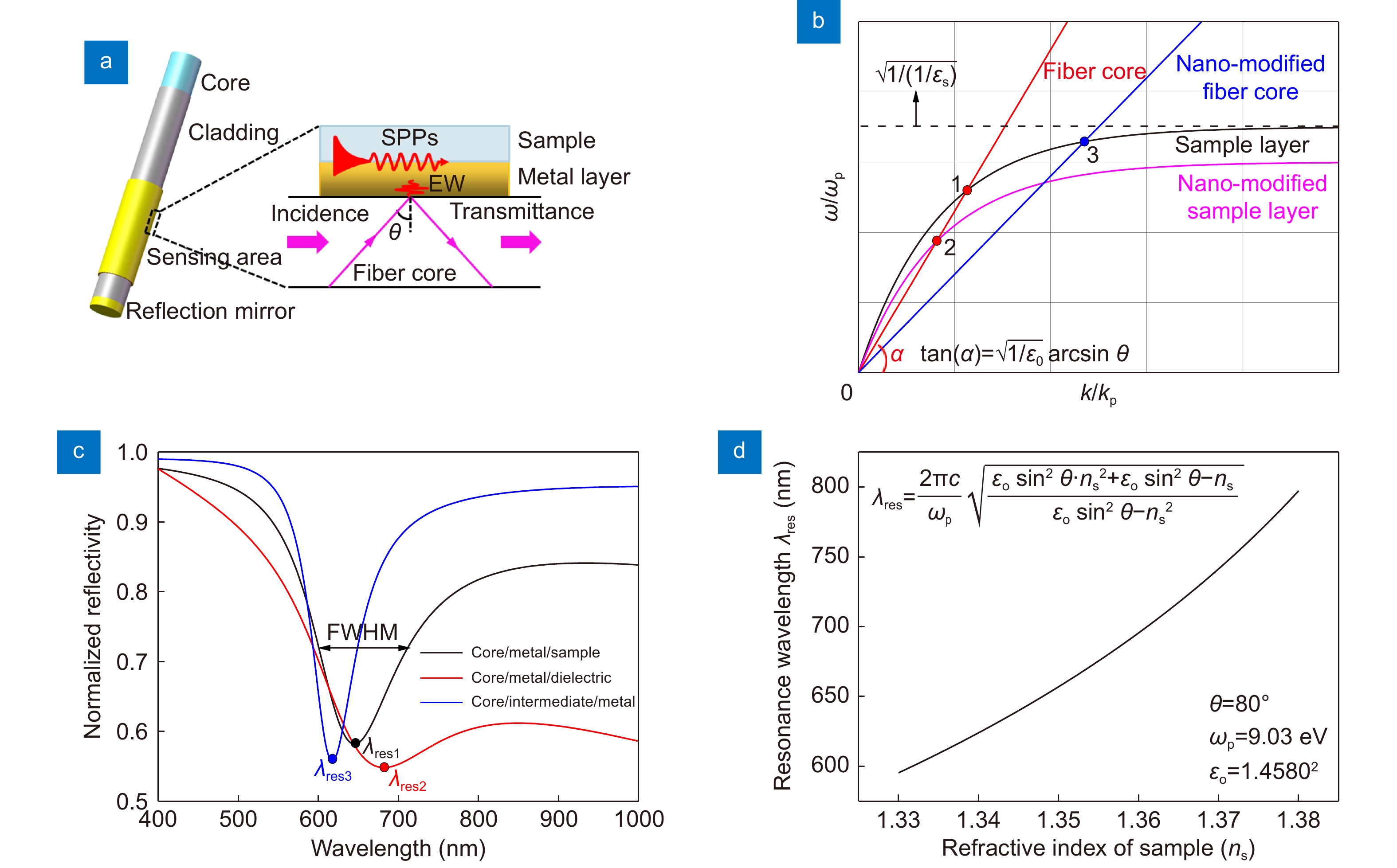
(a) Schematic of the sensing structure of the conventional fiber plasmonic resonance sensor. (b) The dispersion model for configuring plasmonic resonance spectral characteristics. (c) Plasmonic resonance spectra stimulated by different sensing structures. (d) The variation of the resonance wavelength with the RI of the sample layer. ωp is the plasma frequency of Au.
-
Figure 2.
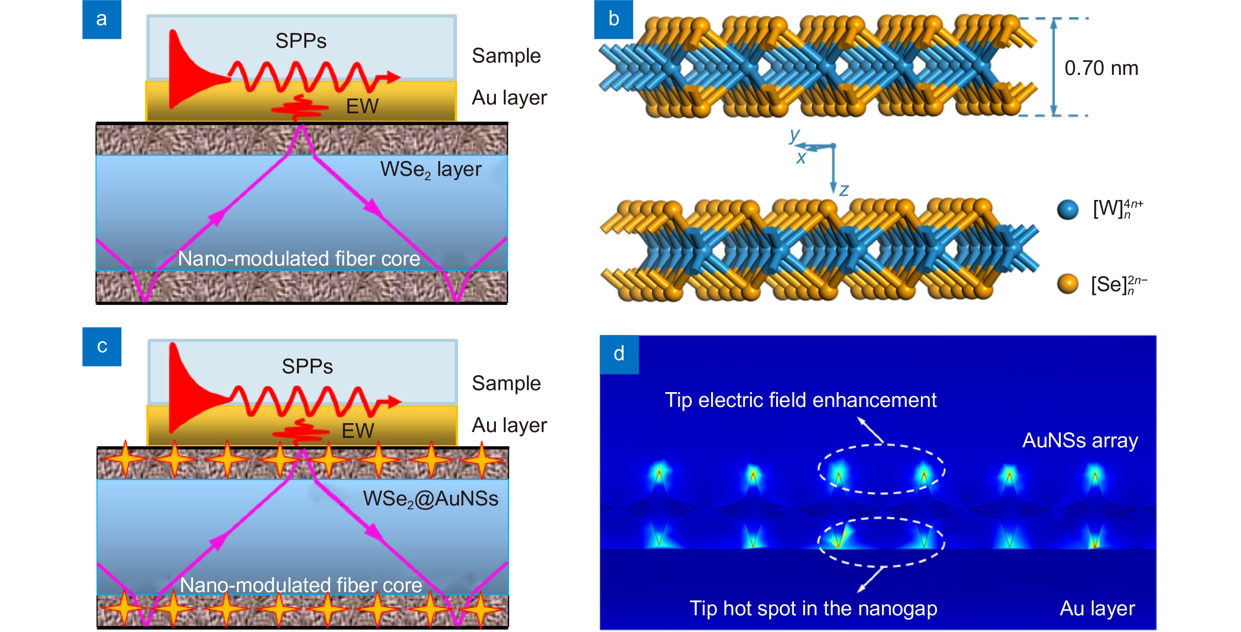
Schematic of the sensing structure of (a) the NMF-CPR sensor and (b) the bilayer structure of WSe2 in the hexagonal crystal system. (c) Schematic of the sensing structure of the tip hot spot enhanced NMF-CPR sensor. (d) The tip electric field enhancement and the tip hot spot in the nanogap between AuNSs and the Au layer.
-
Figure 3.
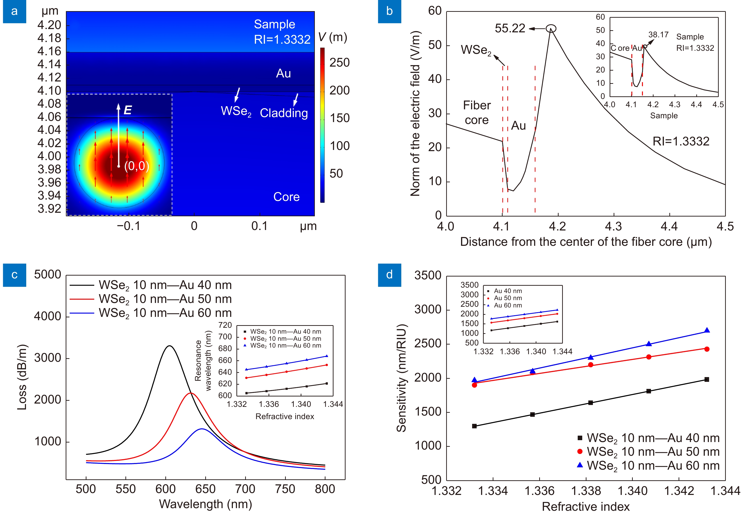
(a) The localized mode field distribution of the NMF-CPR sensor with a WSe2 layer of 10 nm and an Au layer of 50 nm. Inset: the overall mode field distribution around the fiber core. (b) The vertical electric field distribution of the NMF-CPR sensor. Inset: the electric field distribution of the conventional fiber plasmonic resonance sensor with an Au layer of 50 nm. (c) Initial loss peaks of NMF-CPR sensors with different thicknesses of Au layers. Inset: binomial fitting curves of resonance wavelengths and RI points. The tangent slope of each point on the curve represents the sensitivity of the sensor at the corresponding RI point. (d) The sensitivity of NMF-CPR sensors with different thicknesses of Au layers corresponding to each RI. Inset: the sensitivity of conventional fiber plasmonic resonance sensors with different thicknesses of Au layers.
-
Figure 4.
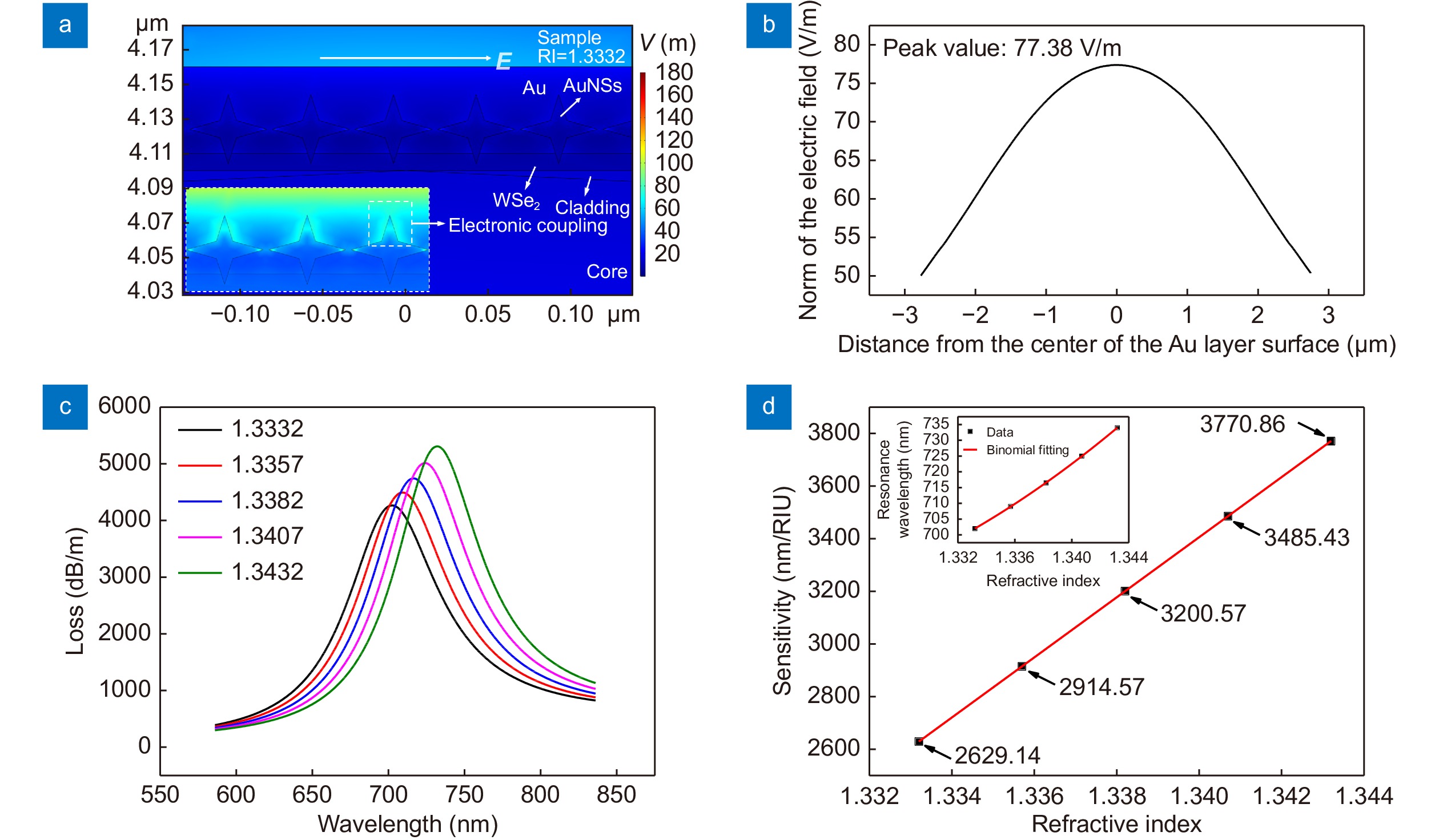
(a) The mode field distribution, (b) the horizontal electric field distribution on the upper surface, (c) loss spectra and (d) the sensitivity at each RI point of the tip hot spot enhanced NMF-CPR sensor with a WSe2 layer of 10 nm, AuNSs of 40 nm, and an Au layer of 50 nm. The inset in (a) is an enlarged view of the mode field distribution around AuNSs. The color legend has been adjusted so that the electronic coupling is more clearly represented. The inset in (d) is the binomial fitting curve of resonance wavelengths and RI points. The tangent slope of each point on the curve represents the sensitivity of the sensor at the corresponding RI point.
-
Figure 5.
(a) Initial resonance dips of NMF-CPR sensors with different thicknesses of Au layers. Inset: binomial fitting curves of resonance wavelengths and RI points. The tangent slope of each point on the curve represents the sensitivity of the sensor at the corresponding RI point. (b) The sensitivity of NMF-CPR sensors with different thicknesses of Au layers corresponding to each RI. Inset: the sensitivity of conventional fiber plasmonic resonance sensors with different thicknesses of Au layers.
-
Figure 6.
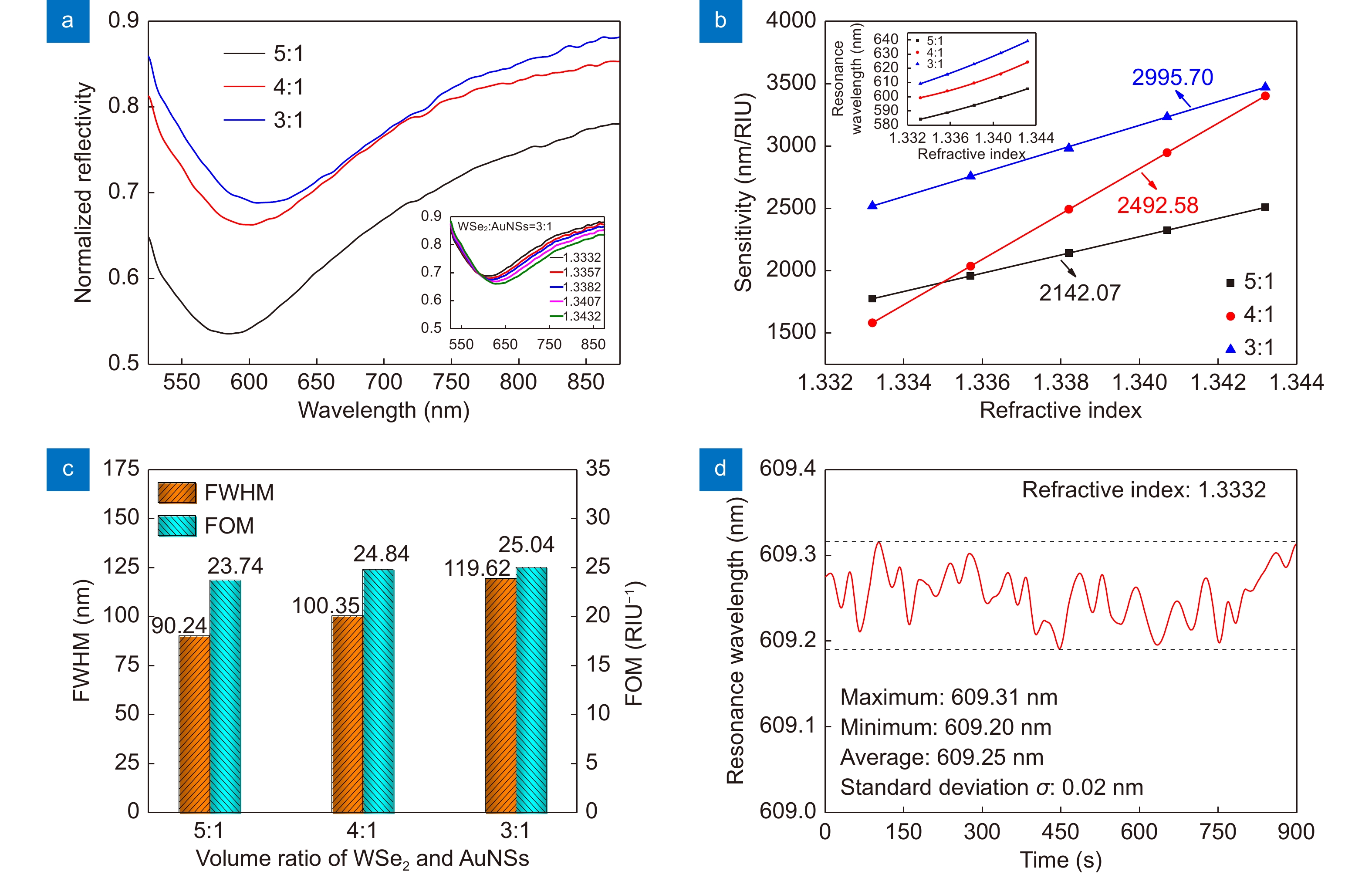
(a) Initial resonance dips of tip hot spot enhanced NMF-CPR sensors with different doping proportions (WSe2∶AuNSs). Inset: resonance spectra of the sensor with a doping proportion of 3∶1. (b) The sensitivity of tip hot spot enhanced NMF-CPR sensors with different doping proportions corresponding to each RI. The average sensitivity of the sensors is 2142.07 nm/RIU, 2492.58 nm/RIU and 2995.70 nm/RIU, respectively. Inset: binomial fitting curves of resonance wavelengths and RI points. The tangent slope of each point on the curve represents the sensitivity of the sensor at the corresponding RI point. (c) The FWHM of initial resonance dips and the FOM of the sensors with different doping proportions. (d) Monitoring of the resonance wavelength of the tip hot spot enhanced NMF-CPR sensor with a doping proportion of 3∶1.

 E-mail Alert
 RSS
RSS


 DownLoad:
