
 E-mail Alert
 RSS
RSS
| Citation: |
Wei X R, Liang Y Z, He Y J, et al. Tamm-surface plasmon hybrid mode for improving sensing figure of merit[J]. Opto-Electron Eng, 2022, 49(11): 220217. doi: 10.12086/oee.2022.220217

|
Tamm-surface plasmon hybrid mode for improving sensing figure of merit
-
Abstract
The hybrid coupling of Tamm plasmon polariton (TPP) and surface plasmon polariton (SPP) on the surface of a gold film based on the prism coupling has attracted extensive attention and has been widely investigated. However, the traditional excitation configuration has bulky optical elements and requires accurate control of the angle of incident light, which limits its integration and practical application. In order to simplify the excitation condition of the TPP-SPP hybrid mode, a feasible grating-coupled multilayer stack structure is proposed in this paper. The structure mainly consists of three parts: a nanometric thin gold film on the top layer, a one-dimensional Bragg photonic crystal in the middle, and a gold nanograting on the bottom. In this structure, the SPR and TPP resonance excitations on the upper and lower surfaces of the top gold film are simultaneously achieved by utilizing the first-order transmitted light of the bottom nanograting. The hybrid coupling between the two modes greatly reduces the resonance bandwidth of the generated mode, thereby significantly improving the sensing figure of merit of the generated mode. Additionally, the hybrid coupling of both SPP and TPP modes can be realized in a wide spectral range by altering the period of the nanograting and the thickness of the one-dimensional Bragg photonic crystal. Compared with the traditional prism-coupled TPP and SPP dual-mode coupling structures, the designed grating-coupled multilayer nanostructure can realize the resonant coupling of the two modes at the normal incidence without prism and limitation of incident angle. This not only facilitates the further integration and miniaturization of the structure, but also has important significance for broadening the practical application of surface plasmon resonance sensors. -

-
References
[1] Singh P. SPR biosensors: historical perspectives and current challenges[J]. Sens Actuators B Chem, 2016, 229: 110−130. doi: 10.1016/j.snb.2016.01.118 [2] Wen X M, Bi Y G, Yi F S, et al. Tunable surface plasmon-polariton resonance in organic light-emitting devices based on corrugated alloy electrodes[J]. Opto-Electron Adv, 2021, 4(8): 200024. doi: 10.29026/oea.2021.200024 [3] Hu A Q, Liu S, Zhao J Y, et al. Controlling plasmon‐exciton interactions through photothermal reshaping[J]. Opto-Electron Adv, 2020, 3(1): 190017. doi: 10.29026/oea.2020.190017 [4] Tong L M, Wei H, Zhang S P, et al. Recent advances in plasmonic sensors[J]. Sensors, 2014, 14(5): 7959−7973. doi: 10.3390/s140507959 [5] Zhao Y, Tong R J, Xia F, et al. Current status of optical fiber biosensor based on surface plasmon resonance[J]. Biosens Bioelectron, 2019, 142: 111505. doi: 10.1016/j.bios.2019.111505 [6] Shrivastav A M, Cvelbar U, Abdulhalim I. A comprehensive review on plasmonic-based biosensors used in viral diagnostics[J]. Commun Biol, 2021, 4(1): 70. doi: 10.1038/s42003-020-01615-8 [7] Verma R, Gupta B D. Detection of heavy metal ions in contaminated water by surface plasmon resonance based optical fibre sensor using conducting polymer and chitosan[J]. Food Chem, 2015, 166: 568−575. doi: 10.1016/j.foodchem.2014.06.045 [8] Li Y, Liu X, Lin Z. Recent developments and applications of surface plasmon resonance biosensors for the detection of mycotoxins in foodstuffs[J]. Food Chem, 2012, 132(3): 1549−1554. doi: 10.1016/j.foodchem.2011.10.109 [9] Homola J, Yee S S, Gauglitz G. Surface plasmon resonance sensors: review[J]. Sens Actuators B Chem, 1999, 54(1–2): 3–15. doi: 10.1016/S0925-4005(98)00321-9. [10] Kasani S, Curtin K, Wu N Q. A review of 2D and 3D plasmonic nanostructure array patterns: fabrication, light management and sensing applications[J]. Nanophotonics, 2019, 8(12): 2065−2089. doi: 10.1515/nanoph-2019-0158 [11] Li D C, Wu J W, Wu P, et al. Affinity based glucose measurement using fiber optic surface plasmon resonance sensor with surface modification by borate polymer[J]. Sens Actuators B Chem, 2015, 213: 295−304. doi: 10.1016/j.snb.2015.02.039 [12] Wu L M, Guo J, Wang Q K, et al. Sensitivity enhancement by using few-layer black phosphorus-graphene/TMDCs heterostructure in surface plasmon resonance biochemical sensor[J]. Sens Actuators B Chem, 2017, 249: 542−548. doi: 10.1016/j.snb.2017.04.110 [13] Li C, Guo J J, Shafi M, et al. Optical fiber SPR biosensor complying with a 3D composite hyperbolic metamaterial and a graphene film[J]. Photonics Res, 2021, 9(3): 379−388. doi: 10.1364/PRJ.416815 [14] Kumar S, Maji P S, Das R. Tamm-plasmon resonance based temperature sensor in a Ta2O5/SiO2 based distributed Bragg reflector[J]. Sens Actuators A Phys, 2017, 260: 10−15. doi: 10.1016/j.sna.2017.03.038 [15] Wang X, Jiang X, You Q, et al. Tunable and multichannel terahertz perfect absorber due to Tamm surface plasmons with graphene[J]. Photonics Res, 2017, 5(6): 536−542. doi: 10.1364/PRJ.5.000536 [16] Chang C C, Chen T Y, Lin T W, et al. Flexible and ultranarrow transmissive color filters by simultaneous excitations of triple resonant eigenmodes in hybrid metallic–optical Tamm state devices[J]. ACS Photonics, 2021, 8(2): 540−549. doi: 10.1021/acsphotonics.0c01550 [17] Buzavaite-Verteliene E, Plikusiene I, Tolenis T, et al. Hybrid Tamm-surface plasmon polariton mode for highly sensitive detection of protein interactions[J]. Opt Express, 2020, 28(20): 29033−29043. doi: 10.1364/OE.401802 [18] Afinogenov B I, Bessonov V O, Nikulin A A, et al. Observation of hybrid state of Tamm and surface plasmon-polaritons in one-dimensional photonic crystals[J]. Appl Phys Lett, 2013, 103(6): 061112. doi: 10.1063/1.4817999 [19] Bužavaitė-Vertelienė E, Vertelis V, Balevičius Z. The experimental evidence of a strong coupling regime in the hybrid Tamm plasmon-surface plasmon polariton mode[J]. Nanophotonics, 2021, 10(5): 1565−1571. doi: 10.1515/nanoph-2020-0660 [20] He X L, Yi H, Long J, et al. Plasmonic crystal cavity on single-mode optical fiber end facet for label-free biosensing[J]. Appl Phys Lett, 2016, 108(23): 231105. doi: 10.1063/1.4953413 [21] Lei Z Y, Zhou X, Yang J, et al. Second-order distributed-feedback surface plasmon resonator for single-mode fiber end-facet biosensing[J]. Appl Phys Lett, 2017, 110(17): 171107. doi: 10.1063/1.4982625 [22] Johnson P B, Christy R W. Optical constants of the noble metals[J]. Phys Rev B, 1972, 6(12): 4370−4379. doi: 10.1103/PhysRevB.6.4370 [23] 侯艺博, 霍义萍, 姜雪莹, 等. 新月十字架纳米结构中多Fano共振的产生和高FOM共振(英文)[J]. 光电工程, 2020, 47(11): 200010. doi: 10.12086/oee.2020.200010. Hou Y B, Huo Y P, Jiang X Y, et al. Generation of multiple Fano resonance and high FOM resonance based on the crescent cross nanostructure[J]. Opto-Electron Eng, 2020, 47(11): 200010. doi: 10.12086/oee.2020.200010. -
Overview
Surface plasmon resonance (SPR) sensing technology has attracted widespread attention due to its advantages of high sensitivity, label-free, and real-time dynamic monitoring. Traditional SPR sensing platform needs the use of a prism, and requires that the transverse magnetic (TM) polarized light incident at a specific angle to satisfy the wave vector matching condition and excite the surface plasmon polariton (SPP) mode at the interface between the metal film and the external environment. Moreover, Tamm plasmon polariton (TPP), as a special plasmon boundary state mode, can be excited by using the boundary between the one-dimensional Bragg photonic crystal (PC) and the metal film and has broad application prospects in the fields of new optoelectronic devices. Compared with SPP, the excitation of TPP does not require wavevector compensation for incident light and can be achieved at any polarization. However, the enhanced electromagnetic field of the TPP mode is mainly localized inside the structure and cannot sense the changes in the external environment, which greatly limits its application in the field of biochemical sensing. To break through this limitation, researchers integrated the one-dimensional Bragg PC structures onto the traditional prism structures to achieve hybrid coupling of SPP mode and TPP mode by using the oblique incident light, which could improve the sensing performance of the SPR sensors. However, this kind of TPP-SPP strong coupling excitation also requires a bulky prism and a precise incident light angle control system, which is not conducive to the miniaturization and integrated application of the structure. Therefore, we propose a feasible design of a grating-coupled multilayer stack in this paper. The structure mainly consists of three parts: a nanometric gold film on the top layer, a one-dimensional Bragg PC in the middle, and a gold nanograting on the bottom. In this structure, the SPP and TPP resonance excitations on the upper and lower surfaces of the top nano-gold film are simultaneously achieved by utilizing the first-order transmitted light of the bottom nanograting. The coupling hybridization between the two modes greatly reduces the resonance bandwidth of the generated hybrid mode, resulting in a significant improvement in its sensing figure of merit. In addition, the coupling hybridization of the SPP and the TPP can be realized in a wide spectral range by changing the period of the nanograting and the thickness of the dielectric layers constituting the one-dimensional Bragg PC. Compared with the traditional prism TPP and SPP dual-mode coupling structure, the designed multilayer nanostructure can realize the resonance coupling of the two modes over broad wavelength ranges at the normal incidence. These results not only make it easier to further integrate and miniaturize the structure, but also have important significance for broadening the practical application of the surface plasmon resonance sensors.
-
Access History


Export File
Citation
Wei X R, Liang Y Z, He Y J, et al. Tamm-surface plasmon hybrid mode for improving sensing figure of merit[J]. Opto-Electron Eng, 2022, 49(11): 220217. doi: 10.12086/oee.2022.220217
Format
Content
 DownLoad:


-
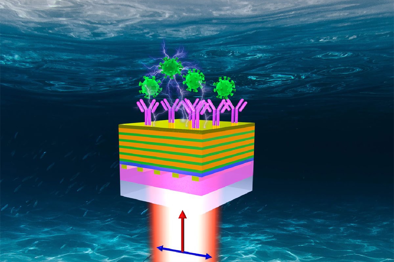
Figure 1.
Nanograting coupled multilayer stack structure for improving sensing figure of merit. (a) Three-dimensional schematic for biomolecule detection; (b) Cross-sectional view of the structure and corresponding material composition. The red arrows represent the propagation path of the TM polarized light, and the red curves represent the distribution characteristics of the electric field of SPP and TPP in the structure
-
Figure 2.
The reflection spectra of the simplified structure. When the incident angle is equal to the first-order diffraction angle of the nanograting with a period of 536 nm and the thicknesses of the gold film on the top surface of the structure are (a) 400 nm and (b) 40 nm, respectively, the effect of the center wavelength of the photonic crystal on the reflection spectra of the gold film-photonic crystal multilayer structure; (c) The relationship between the wavevector and the energy of the incident light in the structure, and the dispersion curves excited separately by the SPP mode (yellow short dashed curve) and the low-order TPP mode (cyan short dashed line) ; (d) The reflection spectra of the structure that only excites TPP mode (yellow curve) and SPP mode (blue curve), and the reflection spectra corresponding to the strong coupling between TPP and SPP (red curve)
-
Figure 3.
Reflection spectra and electric field distributions at the resonance positions of the grating-coupled multilayer stack structure. (a) Reflection spectra of three typical structures, in which there are TPP (yellow curve) and SPP (blue curve) modes and TPP-SPP hybrid mode (red curve) generated; The spatial electric field intensity distributions of (b) SPP mode, (c) TPP mode; (d) High-frequency and (e) low-frequency TPP-SPP hybridization modes. The curve in the right inset is the variation of electric field intensity at the dotted line position in the electric field intensity distribution diagrams
-
Figure 4.
Quantitative evaluation of the sensing performance. Reflection spectra of structures with (a) TPP-SPP hybrid mode and (b) SPP mode at the ambient with different refractive indexes; (c) The amount of red-shift of the resonance wavelengths caused by the change of the external refractive index; (d) Sensing figures of merit of TPP-SPP hybrid mode (red mark) and SPP mode (blue mark) under the ambient surroundings with different refractive indexes
-
Figure 5.
Reflection spectra of the structures corresponding to the excited high-frequency TPP-SPP hybrid mode structures at the wavelengths of (a) 631 nm and (b) 844 nm. The reflection spectra of the SPP mode are used as a reference; (c) Bulk refractive index sensitivity and (d) sensing figure of merit of TPP-SPP hybrid mode and SPP mode at different structural periods
- Figure .


