| Citation: | Wu GX, Zhu RZ, Lu YQ et al. Optical scanning endoscope via a single multimode optical fiber. Opto-Electron Sci 3, 230041 (2024). doi: 10.29026/oes.2024.230041 |
-
Abstract
Optical endoscopy has become an essential diagnostic and therapeutic approach in modern biomedicine for directly observing organs and tissues deep inside the human body, enabling non-invasive, rapid diagnosis and treatment. Optical fiber endoscopy is highly competitive among various endoscopic imaging techniques due to its high flexibility, compact structure, excellent resolution, and resistance to electromagnetic interference. Over the past decade, endoscopes based on a single multimode optical fiber (MMF) have attracted widespread research interest due to their potential to significantly reduce the footprint of optical fiber endoscopes and enhance imaging capabilities. In comparison with other imaging principles of MMF endoscopes, the scanning imaging method based on the wavefront shaping technique is highly developed and provides benefits including excellent imaging contrast, broad applicability to complex imaging scenarios, and good compatibility with various well-established scanning imaging modalities. In this review, various technical routes to achieve light focusing through MMF and procedures to conduct the scanning imaging of MMF endoscopes are introduced. The advancements in imaging performance enhancements, integrations of various imaging modalities with MMF scanning endoscopes, and applications are summarized. Challenges specific to this endoscopic imaging technology are analyzed, and potential remedies and avenues for future developments are discussed.-
Keywords:
- multimode optical fiber /
- endoscope /
- scanning imaging /
- focusing /
- wavefront shaping
-

-
References
[1] Litynski GS. Endoscopic surgery: the history, the pioneers. World J Surg 23, 745–753 (1999). doi: 10.1007/s002689900576 [2] Vasquez-Lopez SA, Turcotte R, Koren V et al. Subcellular spatial resolution achieved for deep-brain imaging in vivo using a minimally invasive multimode fiber. Light Sci Appl 7, 110 (2018). doi: 10.1038/s41377-018-0111-0 [3] Gaab MR. Instrumentation: endoscopes and equipment. World Neurosurg 79, S14.e11–S14.e21 (2013). doi: 10.1016/j.wneu.2012.02.032 [4] Liu HH, Hu DJJ, Sun QZ et al. Specialty optical fibers for advanced sensing applications. Opto-Electron Sci 2, 220025 (2023). doi: 10.29026/oes.2023.220025 [5] Jiang BQ, Hou YG, Wu JX et al. In-fiber photoelectric device based on graphene-coated tilted fiber grating. Opto-Electron Sci 2, 230012 (2023). doi: 10.29026/oes.2023.230012 [6] Hopkins HH, Kapany NS. A flexible fibrescope, using static scanning. Nature 173, 39–41 (1954). [7] Hopkins HH, Kapany NS. Transparent fibres for the transmission of optical images. Opt Acta Int J Opt 1, 164–170 (1955). doi: 10.1080/713818685 [8] Kao KC, Hockham GA. Dielectric-fibre surface waveguides for optical frequencies. Proc Inst Electr Eng 113, 1151–1158 (1966). doi: 10.1049/piee.1966.0189 [9] Sun JW, Wu JC, Wu S et al. Quantitative phase imaging through an ultra-thin lensless fiber endoscope. Light Sci Appl 11, 204 (2022). doi: 10.1038/s41377-022-00898-2 [10] Orth A, Ploschner M, Wilson E et al. Optical fiber bundles: ultra-slim light field imaging probes. Sci Adv 5, eaav1555 (2019). doi: 10.1126/sciadv.aav1555 [11] Pan YT, Xie HK, Fedder GK. Endoscopic optical coherence tomography based on a microelectromechanical mirror. Opt Lett 26, 1966–1968 (2001). doi: 10.1364/OL.26.001966 [12] Smithwick QYJ, Seibel EJ, Reinhall PG et al. Control aspects of the single-fiber scanning endoscope. Proc SPIE 4253, 176–188 (2001). doi: 10.1117/12.427920 [13] Liu XM, Cobb MJ, Chen YC et al. Rapid-scanning forward-imaging miniature endoscope for real-time optical coherence tomography. Opt Lett 29, 1763–1765 (2004). doi: 10.1364/OL.29.001763 [14] Kaur M, Lane PM, Menon C. Scanning and actuation techniques for cantilever-based fiber optic endoscopic scanners—a review. Sensors 21, 251 (2021). doi: 10.3390/s21010251 [15] Kang JQ, Zhu R, Sun YX et al. Pencil-beam scanning catheter for intracoronary optical coherence tomography. Opto-Electron Adv 5 5, 200050 (2022). doi: 10.29026/oea.2022.200050 [16] Hirschowitz BI, Curtiss LE, Peters CW et al. Demonstration of a new gastroscope, the “fiberscope”. Gastroenterology 35, 50–53 (1958). doi: 10.1016/S0016-5085(19)35579-9 [17] Yan XQ, Li M, Chen ZQ et al. Schlemm’s canal and trabecular meshwork in eyes with primary open angle glaucoma: a comparative study using high-frequency ultrasound biomicroscopy. PLoS One 11, e0145824 (2016). doi: 10.1371/journal.pone.0145824 [18] Kaur M, Lane PM, Menon C. Design of scanning fiber micro-cantilever based catheter for ultra-small endoscopes. In Proceedings of 2023 IEEE World AI IoT Congress 717–723 (IEEE, 2023);http://doi.org/10.1109/AIIoT58121.2023.10174425. [19] Chen XP, Reichenbach KL, Xu C. Experimental and theoretical analysis of core-to-core coupling on fiber bundle imaging. Opt Express 16, 21598–21607 (2008). doi: 10.1364/OE.16.021598 [20] Parker HE, Perperidis A, Stone JM et al. Core crosstalk in ordered imaging fiber bundles. Opt Lett 45, 6490–6493 (2020). doi: 10.1364/OL.405764 [21] Kyrish M, Kester R, Richards-Kortum R et al. Improving spatial resolution of a fiber bundle optical biopsy system. Proc SPIE 7558, 755807 (2010). doi: 10.1117/12.842744 [22] Papadopoulos IN, Farahi S, Moser C et al. Focusing and scanning light through a multimode optical fiber using digital phase conjugation. Opt Express 20, 10583–10590 (2012). doi: 10.1364/OE.20.010583 [23] Čižmár T, Dholakia K. Shaping the light transmission through a multimode optical fibre: complex transformation analysis and applications in biophotonics. Opt Express 19, 18871–18884 (2011). doi: 10.1364/OE.19.018871 [24] Caravaca-Aguirre AM, Niv E, Conkey DB et al. Real-time resilient focusing through a bending multimode fiber. Opt Express 21, 12881–12887 (2013). doi: 10.1364/OE.21.012881 [25] Čižmár T, Dholakia K. Exploiting multimode waveguides for pure fibre-based imaging. Nat Commun 3, 1027 (2012). doi: 10.1038/ncomms2024 [26] Di Leonardo R, Bianchi S. Hologram transmission through multi-mode optical fibers. Opt Express 19, 247–254 (2011). doi: 10.1364/OE.19.000247 [27] Zhao TR, Ourselin S, Vercauteren T et al. Seeing through multimode fibers with real-valued intensity transmission matrices. Opt Express 28, 20978–20991 (2020). doi: 10.1364/OE.396734 [28] Abraham E, Zhou JX, Liu ZW. Speckle structured illumination endoscopy with enhanced resolution at wide field of view and depth of field. Opto-Electron Adv 6, 220163 (2023). doi: 10.29026/oea.2023.220163 [29] Choi Y, Yoon C, Kim M et al. Scanner-free and wide-field endoscopic imaging by using a single multimode optical fiber. Phys Rev Lett 109, 203901 (2012). doi: 10.1103/PhysRevLett.109.203901 [30] Liu YF, Yu PP, Wu YJ et al. Single-shot wide-field imaging in reflection by using a single multimode fiber. Appl Phys Lett 122, 063701 (2023). doi: 10.1063/5.0132123 [31] Popoff S, Lerosey G, Fink M et al. Image transmission through an opaque material. Nat Commun 1, 81 (2010). doi: 10.1038/ncomms1078 [32] Abrashitova K, Amitonova LV. High-speed label-free multimode-fiber-based compressive imaging beyond the diffraction limit. Opt Express 30, 10456–10469 (2022). doi: 10.1364/OE.444796 [33] Li SH, Saunders C, Lum DJ et al. Compressively sampling the optical transmission matrix of a multimode fibre. Light Sci Appl 10, 88 (2021). doi: 10.1038/s41377-021-00514-9 [34] Amitonova LV, de Boer JF. Compressive imaging through a multimode fiber. Opt Lett 43, 5427–5430 (2018). doi: 10.1364/OL.43.005427 [35] Zhu RZ, Feng HG, Xu F. Deep learning-based multimode fiber imaging in multispectral and multipolarimetric channels. Opt Lasers Eng 161, 107386 (2023). doi: 10.1016/j.optlaseng.2022.107386 [36] Zhu RZ, Luo JX, Zhou XX et al. Anti-perturbation multimode fiber imaging based on the active measurement of the fiber configuration. ACS Photonics 10, 3476–3483 (2023). doi: 10.1021/acsphotonics.3c00390 [37] Xu RC, Zhang LH, Chen ZY et al. High accuracy transmission and recognition of complex images through multimode fibers using deep learning. Laser Photonics Rev 17, 2200339 (2023). doi: 10.1002/lpor.202200339 [38] Wang LL, Yang YS, Liu ZT et al. High‐speed all‐fiber micro‐imaging with large depth of field. Laser Photonics Rev 16, 2100724 (2022). doi: 10.1002/lpor.202100724 [39] Saleh BEA, Teich MC. Fundamentals of Photonics (Wiley, New York, 1991). [40] Xiong W, Hsu CW, Bromberg Y et al. Complete polarization control in multimode fibers with polarization and mode coupling. Light Sci Appl 7, 54 (2018). doi: 10.1038/s41377-018-0047-4 [41] Popoff SM, Lerosey G, Carminati R et al. Measuring the transmission matrix in optics: an approach to the study and control of light propagation in disordered media. Phys Rev Lett 104, 100601 (2010). doi: 10.1103/PhysRevLett.104.100601 [42] Turtaev S, Leite IT, Altwegg-Boussac T et al. High-fidelity multimode fibre-based endoscopy for deep brain in vivo imaging. Light Sci Appl 7, 92 (2018). doi: 10.1038/s41377-018-0094-x [43] Goorden SA, Bertolotti J, Mosk AP. Superpixel-based spatial amplitude and phase modulation using a digital micromirror device. Opt Express 22, 17999–18009 (2014). doi: 10.1364/OE.22.017999 [44] Dubois A, Vabre L, Boccara AC et al. High-resolution full-field optical coherence tomography with a Linnik microscope. Appl Opt 41, 805–812 (2002). doi: 10.1364/AO.41.000805 [45] Bianchi S, Di Leonardo R. A multi-mode fiber probe for holographic micromanipulation and microscopy. Lab Chip 12, 635–639 (2012). doi: 10.1039/C1LC20719A [46] Conkey DB, Caravaca-Aguirre AM, Piestun R. High-speed scattering medium characterization with application to focusing light through turbid media. Opt Express 20, 1733–1740 (2012). doi: 10.1364/OE.20.001733 [47] Ivanina A, Lochocki B, Amitonova LV. Measuring the transmission matrix of a multimode fiber: on-axis vs off-axis holography. Proc SPIE 12574, 125740T (2023). [48] Jákl P, Šiler M, Ježek J et al. Endoscopic imaging using a multimode optical fibre calibrated with multiple internal references. Photonics 9, 37 (2022). doi: 10.3390/photonics9010037 [49] Jákl P, Šiler M, Ježek J et al. Multimode fiber transmission matrix obtained with internal references. Proc SPIE 10886, 1088610 (2019). [50] Collard L, Piscopo L, Pisano F et al. Optimizing the internal phase reference to shape the output of a multimode optical fiber. PLoS One 18, e0290300 (2023). doi: 10.1371/journal.pone.0290300 [51] Amitonova LV, Mosk AP, Pinkse PWH. Rotational memory effect of a multimode fiber. Opt Express 23, 20569–20575 (2015). doi: 10.1364/OE.23.020569 [52] Li SH, Horsley SAR, Tyc T et al. Memory effect assisted imaging through multimode optical fibres. Nat Commun 12, 3751 (2021). doi: 10.1038/s41467-021-23729-1 [53] Drémeau A, Liutkus A, Martina D et al. Reference-less measurement of the transmission matrix of a highly scattering material using a DMD and phase retrieval techniques. Opt Express 23, 11898–11911 (2015). doi: 10.1364/OE.23.011898 [54] N’Gom M, Norris TB, Michielssen E et al. Mode control in a multimode fiber through acquiring its transmission matrix from a reference-less optical system. Opt Lett 43, 419–422 (2018). doi: 10.1364/OL.43.000419 [55] Deng L, Yan JD, Elson DS et al. Characterization of an imaging multimode optical fiber using a digital micro-mirror device based single-beam system. Opt Express 26, 18436–18447 (2018). doi: 10.1364/OE.26.018436 [56] Caramazza P, Moran O, Murray-Smith R et al. Transmission of natural scene images through a multimode fibre. Nat Commun 10, 2029 (2019). doi: 10.1038/s41467-019-10057-8 [57] Huang GQ, Wu DX, Luo JW et al. Retrieving the optical transmission matrix of a multimode fiber using the extended Kalman filter. Opt Express 28, 9487–9500 (2020). doi: 10.1364/OE.389133 [58] Tao XD, Bodington D, Reinig M et al. High-speed scanning interferometric focusing by fast measurement of binary transmission matrix for channel demixing. Opt Express 23, 14168–14187 (2015). doi: 10.1364/OE.23.014168 [59] Zhao TR, Deng L, Wang W et al. Bayes’ theorem-based binary algorithm for fast reference-less calibration of a multimode fiber. Opt Express 26, 20368–20378 (2018). doi: 10.1364/OE.26.020368 [60] Zhao TR, Ourselin S, Vercauteren T et al. Focusing light through multimode fibres using a digital micromirror device: a comparison study of non-holographic approaches. Opt Express 29, 14269–14281 (2021). doi: 10.1364/OE.420718 [61] Plöschner M, Tyc T, Čižmár T. Seeing through chaos in multimode fibres. Nat Photonics 9, 529–535 (2015). doi: 10.1038/nphoton.2015.112 [62] Fischer B, Sternklar S. Image transmission and interferometry with multimode fibers using self‐pumped phase conjugation. Appl Phys Lett 46, 113–114 (1985). doi: 10.1063/1.95703 [63] McMichael I, Yeh P, Beckwith P. Correction of polarization and modal scrambling in multimode fibers by phase conjugation. Opt Lett 12, 507–509 (1987). doi: 10.1364/OL.12.000507 [64] Son JY, Bobrinev VI, Jeon HW et al. Direct image transmission through a multimode optical fiber. Appl Opt 35, 273–277 (1996). doi: 10.1364/AO.35.000273 [65] Dunning GJ, Lind RC. Demonstration of image transmission through fibers by optical phase conjugation. Opt Lett 7, 558–560 (1982). doi: 10.1364/OL.7.000558 [66] Ogasawara T, Ohno M, Karaki K et al. Image transmission with a pair of graded-index optical fibers and a BaTiO3 phase-conjugate mirror. J Opt Soc Am B 13, 2193–2197 (1996). doi: 10.1364/JOSAB.13.002193 [67] Papadopoulos IN, Farahi S, Moser C et al. Focused light delivery and all optical scanning from a multimode optical fiber using digital phase conjugation. Proc SPIE 8576, 857603 (2013). doi: 10.1117/12.2001605 [68] Zhang JW, Ma CJ, Dai SQ et al. Transmission and total internal reflection integrated digital holographic microscopy. Opt Lett 41, 3844–3847 (2016). doi: 10.1364/OL.41.003844 [69] Ma CJ, Di JL, Li Y et al. Rotational scanning and multiple-spot focusing through a multimode fiber based on digital optical phase conjugation. Appl Phys Express 11, 062501 (2018). doi: 10.7567/APEX.11.062501 [70] Papadopoulos IN, Farahi S, Moser C et al. High-resolution, lensless endoscope based on digital scanning through a multimode optical fiber. Biomed Opt Express 4, 260–270 (2013). doi: 10.1364/BOE.4.000260 [71] Mididoddi CK, Lennon RA, Li SH et al. High-fidelity off-axis digital optical phase conjugation with transmission matrix assisted calibration. Opt Express 28, 34692–34705 (2020). doi: 10.1364/OE.409226 [72] Jang M, Ruan HW, Zhou HJ et al. Method for auto-alignment of digital optical phase conjugation systems based on digital propagation. Opt Express 22, 14054–14071 (2014). doi: 10.1364/OE.22.014054 [73] Mahalati RN, Askarov D, Wilde JP et al. Adaptive control of input field to achieve desired output intensity profile in multimode fiber with random mode coupling. Opt Express 20, 14321–14337 (2012). doi: 10.1364/OE.20.014321 [74] Zhao HC, Ma HT, Zhou P et al. Multimode fiber laser beam cleanup based on stochastic parallel gradient descent algorithm. Opt Commun 284, 613–615 (2011). doi: 10.1016/j.optcom.2010.09.039 [75] Yu H, Yao ZY, Sui XB et al. Focusing through disturbed multimode optical fiber based on self-adaptive genetic algorithm. Optik 261, 169129 (2022). doi: 10.1016/j.ijleo.2022.169129 [76] Cheng SF, Zhong TT, Woo CM et al. Long-distance pattern projection through an unfixed multimode fiber with natural evolution strategy-based wavefront shaping. Opt Express 30, 32565–32576 (2022). doi: 10.1364/OE.462275 [77] Li BQ, Zhang B, Feng Q et al. Shaping the wavefront of incident light with a strong robustness particle swarm optimization algorithm. Chin Phys Lett 35, 124201 (2018). doi: 10.1088/0256-307X/35/12/124201 [78] Conkey DB, Brown AN, Caravaca-Aguirre AM et al. Genetic algorithm optimization for focusing through turbid media in noisy environments. Opt Express 20, 4840–4849 (2012). doi: 10.1364/OE.20.004840 [79] Yang ZG, Fang LJ, Zhang XC et al. Controlling a scattered field output of light passing through turbid medium using an improved ant colony optimization algorithm. Opt Lasers Eng 144, 106646 (2021). doi: 10.1016/j.optlaseng.2021.106646 [80] Fayyaz Z, Mohammadian N, Reza Rahimi Tabar M et al. A comparative study of optimization algorithms for wavefront shaping. J Innov Opt Health Sci 12, 1942002 (2019). doi: 10.1142/S1793545819420021 [81] Yin Z, Liu GD, Chen FD et al. Fast-forming focused spots through a multimode fiber based on an adaptive parallel coordinate algorithm. Chin Opt Lett 13, 071404 (2015). doi: 10.3788/COL201513.071404 [82] Chen H, Geng Y, Xu CF et al. Efficient light focusing through an MMF based on two-step phase shifting and parallel phase compensating. Appl Opt 58, 7552–7557 (2019). doi: 10.1364/AO.58.007552 [83] Zhang ZK, Kong DP, Geng Y et al. Lensless multimode fiber imaging based on wavefront shaping. Appl Phys Express 14, 092002 (2021). doi: 10.35848/1882-0786/ac19d4 [84] Gomes AD, Turtaev S, Du Y et al. Near perfect focusing through multimode fibres. Opt Express 30, 10645–10663 (2022). doi: 10.1364/OE.452145 [85] Choi Y, Yoon C, Kim M et al. Disorder-mediated enhancement of fiber numerical aperture. Opt Lett 38, 2253–2255 (2013). doi: 10.1364/OL.38.002253 [86] Papadopoulos IN, Farahi S, Moser C et al. Increasing the imaging capabilities of multimode fibers by exploiting the properties of highly scattering media. Opt Lett 38, 2776–2778 (2013). doi: 10.1364/OL.38.002776 [87] Jang H, Yoon C, Chung E et al. Holistic random encoding for imaging through multimode fibers. Opt Express 23, 6705–6721 (2015). doi: 10.1364/OE.23.006705 [88] Bianchi S, Rajamanickam VP, Ferrara L et al. Focusing and imaging with increased numerical apertures through multimode fibers with micro-fabricated optics. Opt Lett 38, 4935–4938 (2013). doi: 10.1364/OL.38.004935 [89] Amitonova LV, Descloux A, Petschulat J et al. High-resolution wavefront shaping with a photonic crystal fiber for multimode fiber imaging. Opt Lett 41, 497–500 (2016). doi: 10.1364/OL.41.000497 [90] Leite IT, Turtaev S, Jiang X et al. Three-dimensional holographic optical manipulation through a high-numerical-aperture soft-glass multimode fibre. Nat Photonics 12, 33–39 (2018). doi: 10.1038/s41566-017-0053-8 [91] Stellinga D, Phillips DB, Mekhail SP et al. Time-of-flight 3D imaging through multimode optical fibers. Science 374, 1395–1399 (2021). doi: 10.1126/science.abl3771 [92] Leite IT, Turtaev S, Boonzajer Flaes DE et al. Observing distant objects with a multimode fiber-based holographic endoscope. APL Photonics 6, 036112 (2021). doi: 10.1063/5.0038367 [93] Lyu ZP, Osnabrugge G, Pinkse PWH et al. Focus quality in raster-scan imaging via a multimode fiber. Appl Opt 61, 4363–4369 (2022). doi: 10.1364/AO.458146 [94] Descloux A, Amitonova LV, Pinkse PWH. Aberrations of the point spread function of a multimode fiber due to partial mode excitation. Opt Express 24, 18501–18512 (2016). doi: 10.1364/OE.24.018501 [95] Velsink MC, Lyu ZY, Pinkse PWH et al. Comparison of round-and square-core fibers for sensing, imaging, and spectroscopy. Opt Express 29, 6523–6531 (2021). doi: 10.1364/OE.417021 [96] Turcotte R, Sutu E, Schmidt CC et al. Deconvolution for multimode fiber imaging: modeling of spatially variant PSF. Biomed Opt Express 11, 4759–4771 (2020). doi: 10.1364/BOE.399983 [97] Turcotte R, Schmidt CC, Emptage NJ et al. Focusing light in biological tissue through a multimode optical fiber: refractive index matching. Opt Lett 44, 2386–2389 (2019). doi: 10.1364/OL.44.002386 [98] Laporte GPJ, Stasio N, Moser C et al. Enhanced resolution in a multimode fiber imaging system. Opt Express 23, 27484–27493 (2015). doi: 10.1364/OE.23.027484 [99] Loterie D, Farahi S, Papadopoulos I et al. Digital confocal microscopy through a multimode fiber. Opt Express 23, 23845–23858 (2015). doi: 10.1364/OE.23.023845 [100] Webb RH. Confocal optical microscopy. Rep Prog Phys 59, 427–471 (1996). doi: 10.1088/0034-4885/59/3/003 [101] Tučková T, Šiler M, Flaes DEB et al. Computational image enhancement of multimode fibre-based holographic endo-microscopy: harnessing the muddy modes. Opt Express 29, 38206–38220 (2021). [102] Turtaev S, Leite IT, Mitchell KJ et al. Comparison of nematic liquid-crystal and DMD based spatial light modulation in complex photonics. Opt Express 25, 29874–29884 (2017). doi: 10.1364/OE.25.029874 [103] Zhang XH, Wen Z, Ma YG et al. High contrast multimode fiber imaging based on wavelength modulation. Appl Opt 59, 6677–6681 (2020). doi: 10.1364/AO.398490 [104] Plöschner M, Straka B, Dholakia K et al. GPU accelerated toolbox for real-time beam-shaping in multimode fibres. Opt Express 22, 2933–2947 (2014). doi: 10.1364/OE.22.002933 [105] Plöschner M, Čižmár T. Compact multimode fiber beam-shaping system based on GPU accelerated digital holography. Opt Lett 40, 197–200 (2015). doi: 10.1364/OL.40.000197 [106] Lee WH. Binary computer-generated holograms. Appl Opt 18, 3661–3669 (1979). doi: 10.1364/AO.18.003661 [107] Stellinga D, Phillip DB, Mekhail S et al. 3D imaging through a single optical fiber. Proc SPIE 12016, 120160M (2022). [108] Turtaev S, Leite IT, Mitchell KJ et al. Exploiting digital micromirror device for holographic micro-endoscopy. Proc SPIE 10932, 1093203 (2019). [109] Turtaev S, Leite IT, Cizmar T. Liquid-crystal and MEMS modulators for beam-shaping through multimode fibre. In Proceedings of 2018 IEEE Photonics Society Summer Topical Meeting Series 207–208 (IEEE, 2018);http://doi.org/10.1109/PHOSST.2018.8456776. [110] Caravaca-Aguirre AM, Piestun R. Single multimode fiber endoscope. Opt Express 25, 1656–1665 (2017). doi: 10.1364/OE.25.001656 [111] Amitonova LV, de Boer JF. Endo-microscopy beyond the Abbe and Nyquist limits. Light Sci Appl 9, 81 (2020). doi: 10.1038/s41377-020-0308-x [112] Zhu RZ, Feng HG, Xiong YF et al. All-fiber reflective single-pixel imaging with long working distance. Opt Laser Technol 158, 108909 (2023). doi: 10.1016/j.optlastec.2022.108909 [113] Wu YC, Rivenson Y, Wang HD et al. Three-dimensional virtual refocusing of fluorescence microscopy images using deep learning. Nat Methods 16, 1323–1331 (2019). doi: 10.1038/s41592-019-0622-5 [114] Wen Z, Dong ZY, Deng QL et al. Single multimode fibre for in vivo light-field-encoded endoscopic imaging. Nat Photonics 17, 679–687 (2023). doi: 10.1038/s41566-023-01240-x [115] Olshansky R. Mode coupling effects in graded-index optical fibers. Appl Opt 14, 935–945 (1975). doi: 10.1364/AO.14.000935 [116] Flaes DEB, Stopka J, Turtaev S et al. Robustness of light-transport processes to bending deformations in graded-index multimode waveguides. Phys Rev Lett 120, 233901 (2018). doi: 10.1103/PhysRevLett.120.233901 [117] Loterie D, Psaltis D, Moser C. Bend translation in multimode fiber imaging. Opt Express 25, 6263–6273 (2017). doi: 10.1364/OE.25.006263 [118] Farahi S, Ziegler D, Papadopoulos IN et al. Dynamic bending compensation while focusing through a multimode fiber. Opt Express 21, 22504–22514 (2013). doi: 10.1364/OE.21.022504 [119] Gu RY, Mahalati RN, Kahn JM. Design of flexible multi-mode fiber endoscope. Opt Express 23, 26905–26918 (2015). doi: 10.1364/OE.23.026905 [120] Gordon GSD, Gataric M, Ramos AGCP et al. Characterizing optical fiber transmission matrices using metasurface reflector stacks for lensless imaging without distal access. Phys Rev X 9, 041050 (2019). [121] Chen HS, Fontaine NK, Ryf R et al. Remote spatio-temporal focusing over multimode fiber enabled by single-ended channel estimation. IEEE J Sel Top Quantum Electron 26, 7701809 (2020). [122] Wen Z, Wang LQ, Zhang XH et al. Fast volumetric fluorescence imaging with multimode fibers. Opt Lett 45, 4931–4934 (2020). doi: 10.1364/OL.398177 [123] Schmidt CC, Turcotte R, Booth MJ et al. Repeated imaging through a multimode optical fiber using adaptive optics. Biomed Opt Express 13, 662–675 (2022). doi: 10.1364/BOE.448277 [124] Rudolf B, Du Y, Turtaev S et al. Thermal stability of wavefront shaping using a DMD as a spatial light modulator. Opt Express 29, 41808–41818 (2021). doi: 10.1364/OE.442284 [125] Zhou Y, Hong MH. Realization of noncontact confocal optical microsphere imaging microscope. Microsc Res Tech 84, 2381–2387 (2021). doi: 10.1002/jemt.23793 [126] Yang XL, Hong MH. Enhancement of axial resolution and image contrast of a confocal microscope by a microsphere working in noncontact mode. Appl Opt 60, 5271–5277 (2021). doi: 10.1364/AO.425028 [127] Loterie D, Goorden SA, Psaltis D et al. Confocal microscopy through a multimode fiber using optical correlation. Opt Lett 40, 5754–5757 (2015). doi: 10.1364/OL.40.005754 [128] Singh S, Labouesse S, Piestun R. Multiview virtual confocal microscopy through a multimode fiber. In Proceedings of 2021 IEEE 18th International Symposium on Biomedical Imaging 584–587 (IEEE, 2021);http://doi.org/10.1109/ISBI48211.2021.9433950. [129] Singh S, Labouesse S, Piestun R. Multiview scattering scanning imaging confocal microscopy through a multimode fiber. IEEE Trans Comput Imaging 9, 159–171 (2023). doi: 10.1109/TCI.2023.3246224 [130] Loterie D, Psaltis D, Moser C. Confocal microscopy via multimode fibers: fluorescence bandwidth. Proc SPIE 9717, 97171C (2016). [131] So PTC, Dong CY, Masters BR et al. Two-photon excitation fluorescence microscopy. Annu Rev Biomed Eng 2, 399–429 (2000). doi: 10.1146/annurev.bioeng.2.1.399 [132] Cao H, Čižmár T, Turtaev S et al. Controlling light propagation in multimode fibers for imaging, spectroscopy, and beyond. Adv Opt Photonics 15, 524–612 (2023). doi: 10.1364/AOP.484298 [133] Xiong W, Ambichl P, Bromberg Y et al. Spatiotemporal control of light transmission through a multimode fiber with strong mode coupling. Phys Rev Lett 117, 053901 (2016). doi: 10.1103/PhysRevLett.117.053901 [134] Xiong W, Hsu CW, Cao H. Long-range spatio-temporal correlations in multimode fibers for pulse delivery. Nat Commun 10, 2973 (2019). doi: 10.1038/s41467-019-10916-4 [135] Morales-Delgado EE, Farahi S, Papadopoulos IN et al. Delivery of focused short pulses through a multimode fiber. Opt Express 23, 9109–9120 (2015). doi: 10.1364/OE.23.009109 [136] Morales-Delgado EE, Psaltis D, Moser C. Two-photon imaging through a multimode fiber. Opt Express 23, 32158–32170 (2015). doi: 10.1364/OE.23.032158 [137] Pikálek T, Trägårdh J, Simpson S et al. Wavelength dependent characterization of a multimode fibre endoscope. Opt Express 27, 28239–28253 (2019). doi: 10.1364/OE.27.028239 [138] Trägårdh J, Pikálek T, Simpson S et al. Towards focusing broad band light through a multimode fiber endoscope. Proc SPIE 10886, 108860J (2019). [139] Turcotte R, Schmidt CC, Booth MJ et al. Volumetric two-photon fluorescence imaging of live neurons using a multimode optical fiber. Opt Lett 45, 6599–6602 (2020). doi: 10.1364/OL.409464 [140] Sivankutty S, Andresen ER, Cossart R et al. Ultra-thin rigid endoscope: two-photon imaging through a graded-index multi-mode fiber. Opt Express 24, 825–841 (2016). doi: 10.1364/OE.24.000825 [141] Velsink MC, Amitonova LV, Pinkse PWH. Spatiotemporal focusing through a multimode fiber via time-domain wavefront shaping. Opt Express 29, 272–290 (2021). doi: 10.1364/OE.412714 [142] Gusachenko I, Chen MZ, Dholakia K. Raman imaging through a single multimode fibre. Opt Express 25, 13782–13798 (2017). doi: 10.1364/OE.25.013782 [143] Gusachenko I, Nylk J, Tello JA et al. Multimode fibre based imaging for optically cleared samples. Biomed Opt Express 8, 5179–5190 (2017). doi: 10.1364/BOE.8.005179 [144] Deng SN, Loterie D, Konstantinou G et al. Raman imaging through multimode sapphire fiber. Opt Express 27, 1090–1098 (2019). doi: 10.1364/OE.27.001090 [145] Zhang C, Aldana-Mendoza JA. Coherent Raman scattering microscopy for chemical imaging of biological systems. J Phys Photonics 3, 032002 (2021). doi: 10.1088/2515-7647/abfd09 [146] Trägårdh J, Pikálek T, Šerý M et al. Label-free CARS microscopy through a multimode fiber endoscope. Opt Express 27, 30055–30066 (2019). doi: 10.1364/OE.27.030055 [147] Pikálek T, Stibůrek M, Simpson S et al. Suppression of the non-linear background in a multimode fibre CARS endoscope. Biomed Opt Express 13, 862–874 (2022). doi: 10.1364/BOE.450375 [148] Cifuentes A, Pikálek T, Ondráčková P et al. Polarization-resolved second-harmonic generation imaging through a multimode fiber. Optica 8, 1065–1074 (2021). doi: 10.1364/OPTICA.430295 [149] Yang LY, Li YP, Fang F et al. Highly sensitive and miniature microfiber-based ultrasound sensor for photoacoustic tomography. Opto-Electron Adv 5, 200076 (2022). doi: 10.29026/oea.2022.200076 [150] Mezil S, Caravaca-Aguirre AM, Zhang EZ et al. Single-shot hybrid photoacoustic-fluorescent microendoscopy through a multimode fiber with wavefront shaping. Biomed Opt Express 11, 5717–5727 (2020). doi: 10.1364/BOE.400686 [151] Zhao TR, Ma MT, Ourselin S et al. Video-rate dual-modal photoacoustic and fluorescence imaging through a multimode fibre towards forward-viewing endomicroscopy. Photoacoustics 25, 100323 (2022). doi: 10.1016/j.pacs.2021.100323 [152] Zhao TR, Pham TT, Baker C et al. Ultrathin, high-speed, all-optical photoacoustic endomicroscopy probe for guiding minimally invasive surgery. Biomed Opt Express 13, 4414–4428 (2022). doi: 10.1364/BOE.463057 [153] Zhao TR, Zhang MJ, Ourselin S et al. Wavefront shaping-assisted forward-viewing photoacoustic endomicroscopy based on a transparent ultrasound sensor. Appl Sci 12, 12619 (2022). doi: 10.3390/app122412619 [154] Zhao TR, Shi MJ, Ourselin S et al. Deep learning boosts the imaging speed of photoacoustic endomicroscopy. Proc SPIE 12379, 123790J (2023). [155] Ohayon S, Caravaca-Aguirre A, Piestun R et al. Minimally invasive multimode optical fiber microendoscope for deep brain fluorescence imaging. Biomed Opt Express 9, 1492–1509 (2018). doi: 10.1364/BOE.9.001492 [156] Kakkava E, Romito M, Conkey DB et al. Selective femtosecond laser ablation via two-photon fluorescence imaging through a multimode fiber. Biomed Opt Express 10, 423–433 (2019). doi: 10.1364/BOE.10.000423 [157] Stibůrek M, Ondráčková P, Tučková T et al. 110 μm thin endo-microscope for deep-brain in vivo observations of neuronal connectivity, activity and blood flow dynamics. Nat Commun 14, 1897 (2023). doi: 10.1038/s41467-023-36889-z [158] Karbasi S, Frazier RJ, Koch KW et al. Image transport through a disordered optical fibre mediated by transverse Anderson localization. Nat Commun 5, 3362 (2014). doi: 10.1038/ncomms4362 [159] Rivenson Y, Göröcs Z, Günaydin H et al. Deep learning microscopy. Optica 4, 1437–1443 (2017). doi: 10.1364/OPTICA.4.001437 [160] Qiao C, Li D, Liu Y et al. Rationalized deep learning super-resolution microscopy for sustained live imaging of rapid subcellular processes. Nat Biotechnol 41, 367–377 (2023). doi: 10.1038/s41587-022-01471-3 -
Access History

Article Metrics
-
Figure 1.
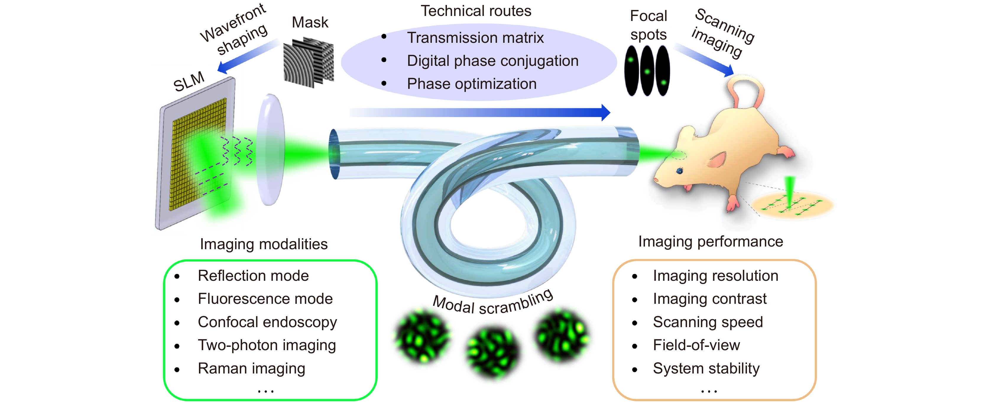
Schematic of MFSE based on wavefront shaping technique. The upper part illustrates three technique routes to acquire appropriate masks for wavefront shaping, aiming at generating high-quality focal spots at the output end of MMF for scanning imaging. The bottom two boxes illustrate the developments of MFSE, including improving imaging performance and combining the MFSE with diverse advanced imaging modalities.
-
Figure 2.
Schematic of optical fields propagating through an MMF. Due to the modal dispersion and mode coupling of the MMF, the input optical field with an ordered profile is converted into a speckled output optical field. ΔβL represents the phase differences between different eigenmodes caused by the intermodal dispersion.
-
Figure 3.
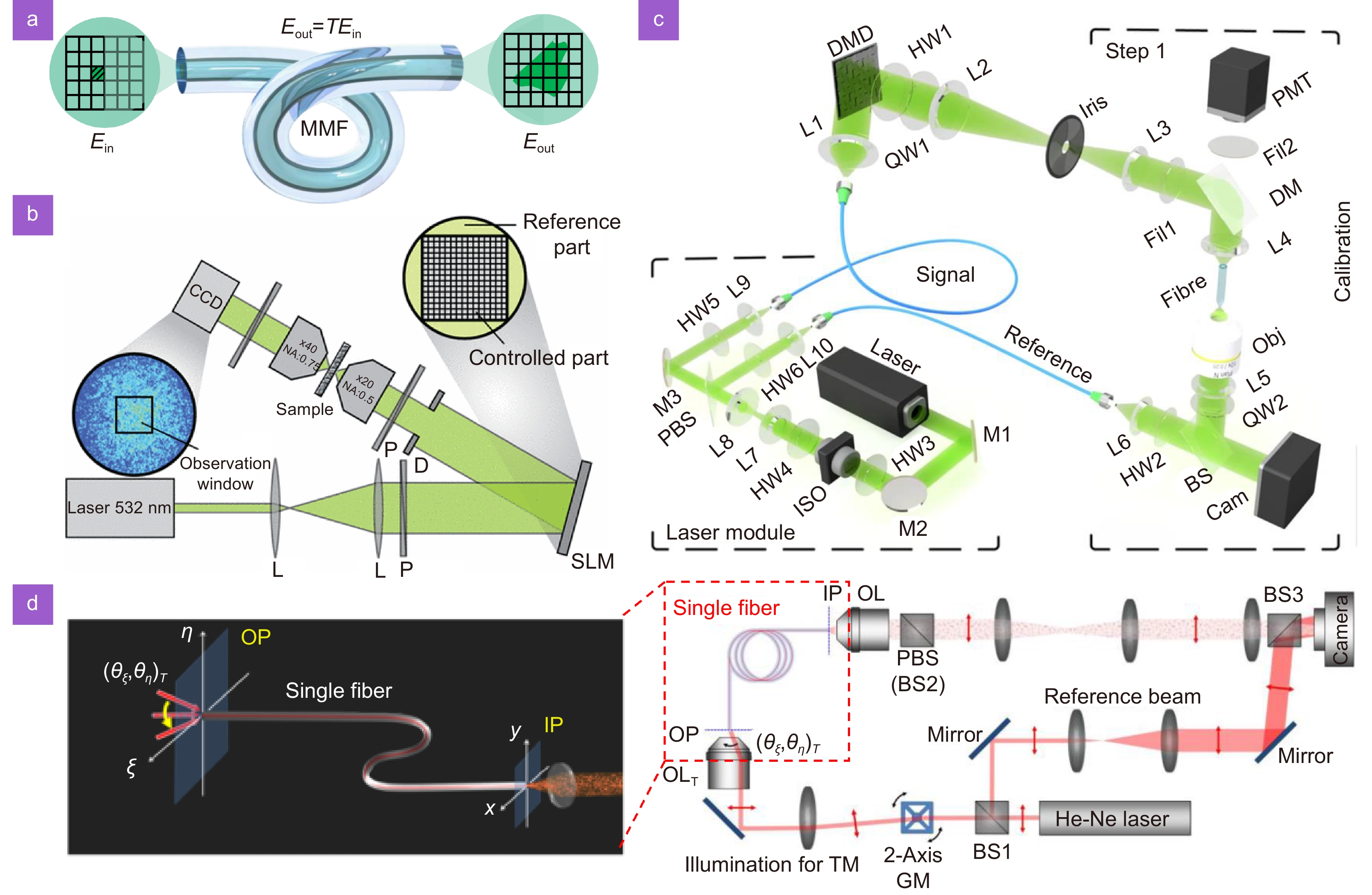
(a) Schematic to illustrate the definition of the transmission matrix of an MMF. Schematics of experimental setups to measure the transmission matrix of MMF or disordered media using (b) internal and (c) external references. (d) Schematic of the experimental setup to measure the transmission matrix through the spatial frequency domain method. Figure reproduced with permission from: (b) ref.41, American Physical Society; (c) ref.42, Springer Nature, under a Creative Commons Attribution 4.0 International License; (d) ref.29, American Physical Society.
-
Figure 4.
(a) Schematic of the experimental setup to implement digital phase conjugation through an MMF, (b) illustration of four steps to realize light focusing through an MMF based on the digital phase conjugation principle. Figure (a) reproduced with permission from ref.22, © Optica Publishing Group.
-
Figure 5.
Principle of the interference-based optimization method. Optimization procedures based on (a−d) internal and (e−g) external reference pathways, respectively. When the external reference pathway is exploited, the effect of polarization on the light focusing can be comprehensively analyzed. Figure reproduced with permission from ref.23, © Optica Publishing Group.
-
Figure 6.
(a) Experimental setup of MFSE works in an all-optical scanning imaging manner. L1-L4: lens, A1: aperture, PD: photodetector, B1: beam splitter or dichroic mirror for reflected light or fluorescence, respectively. (b) Procedures to conduct all-optical scanning imaging, (c) combined image of sequentially generated focal spots at the distal end of MMF and (d) scanning imaging result of a test target. Figures (c) and (d) reproduced with permission from ref.42, Springer Nature, under a Creative Commons Attribution 4.0 International License.
-
Figure 7.
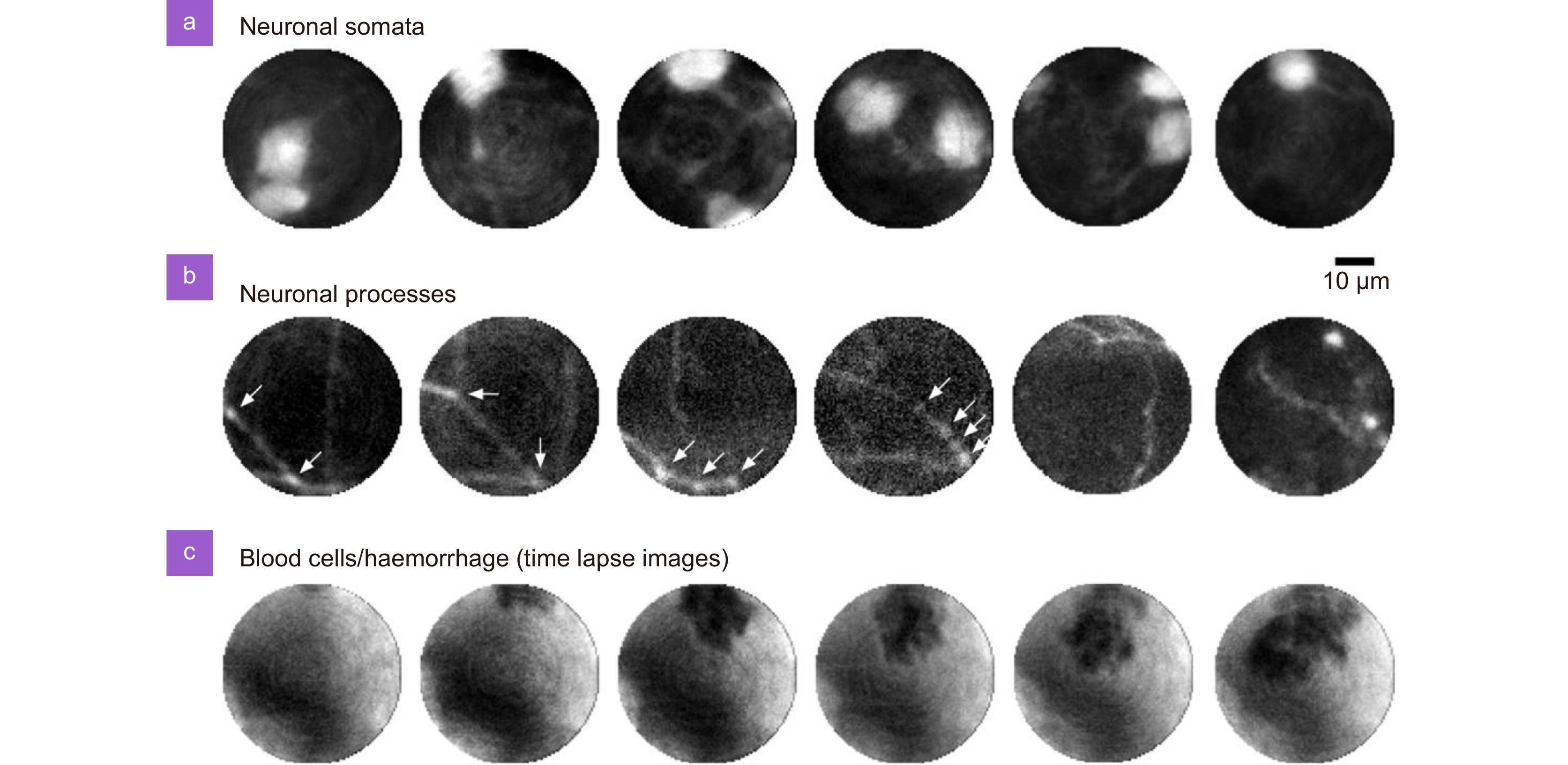
Images of (a) neuronal somata and (b) neuronal processes in the brain of a mouse, as well as (c) a haemorrhage in the primary visual cortex were captured by the MFSE in vivo with an imaging speed of 3.5 frames/s. Figure reproduced with permission from ref.42, Springer Nature, under a Creative Commons Attribution 4.0 International License.
-
Figure 8.
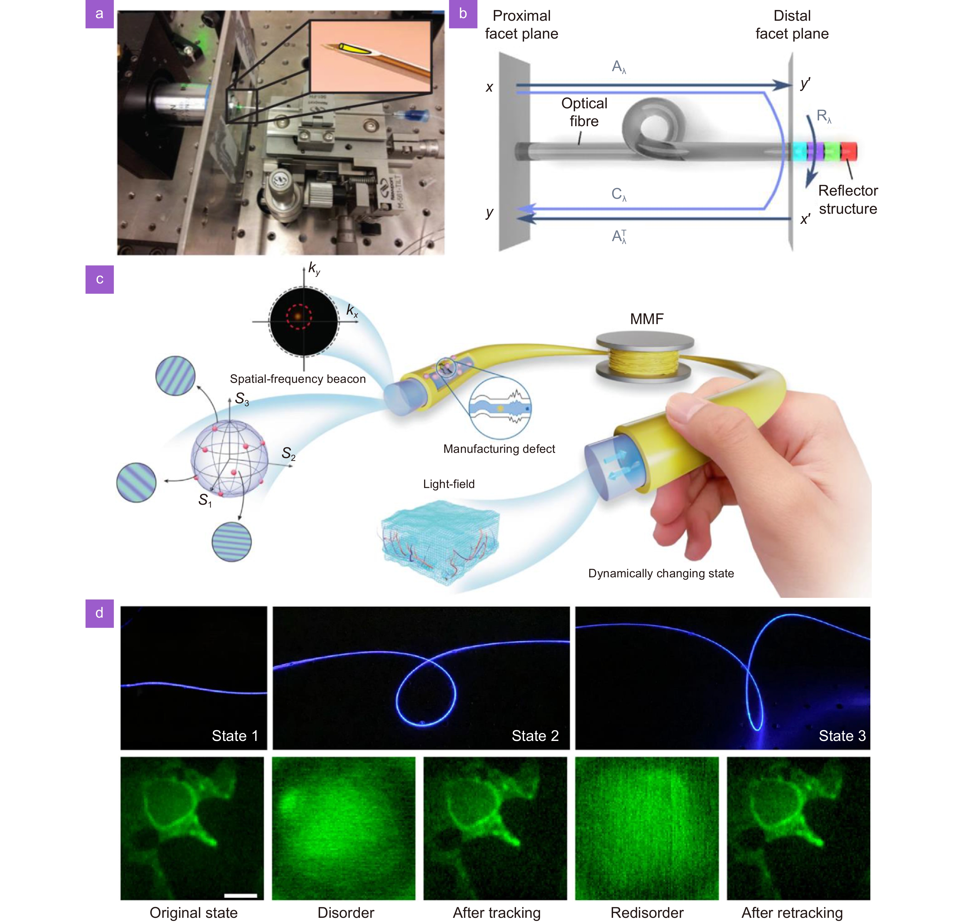
(a) The image of an MMF encapsulated within a 25-gauge needle to prevent the deformation of MMF. (b) Schematic to illustrate the calibration of the transmission matrix at the proximal end of MMF based on the reflection matrix scheme. A metasurface reflector is added at the distal end of MMF to facilitate the measurements of reflection matrices for different wavelengths. (c) Schematic of spatial-frequency tracking adaptive beacon light-field-encoded endoscopy to detect and track disorders caused by movements and manufacturing defects. (d) The geometric state change and the corresponding imaging recovery using the STABLE tracking method. Figure reproduced with permission from: (a) ref.70, © Optica Publishing Group, (b) ref.120, American Physical Society, under a Creative Commons Attribution 4.0 International License, (c) and (d) ref.114, Springer Nature, under a Creative Commons Attribution 4.0 International License.
-
Figure 9.
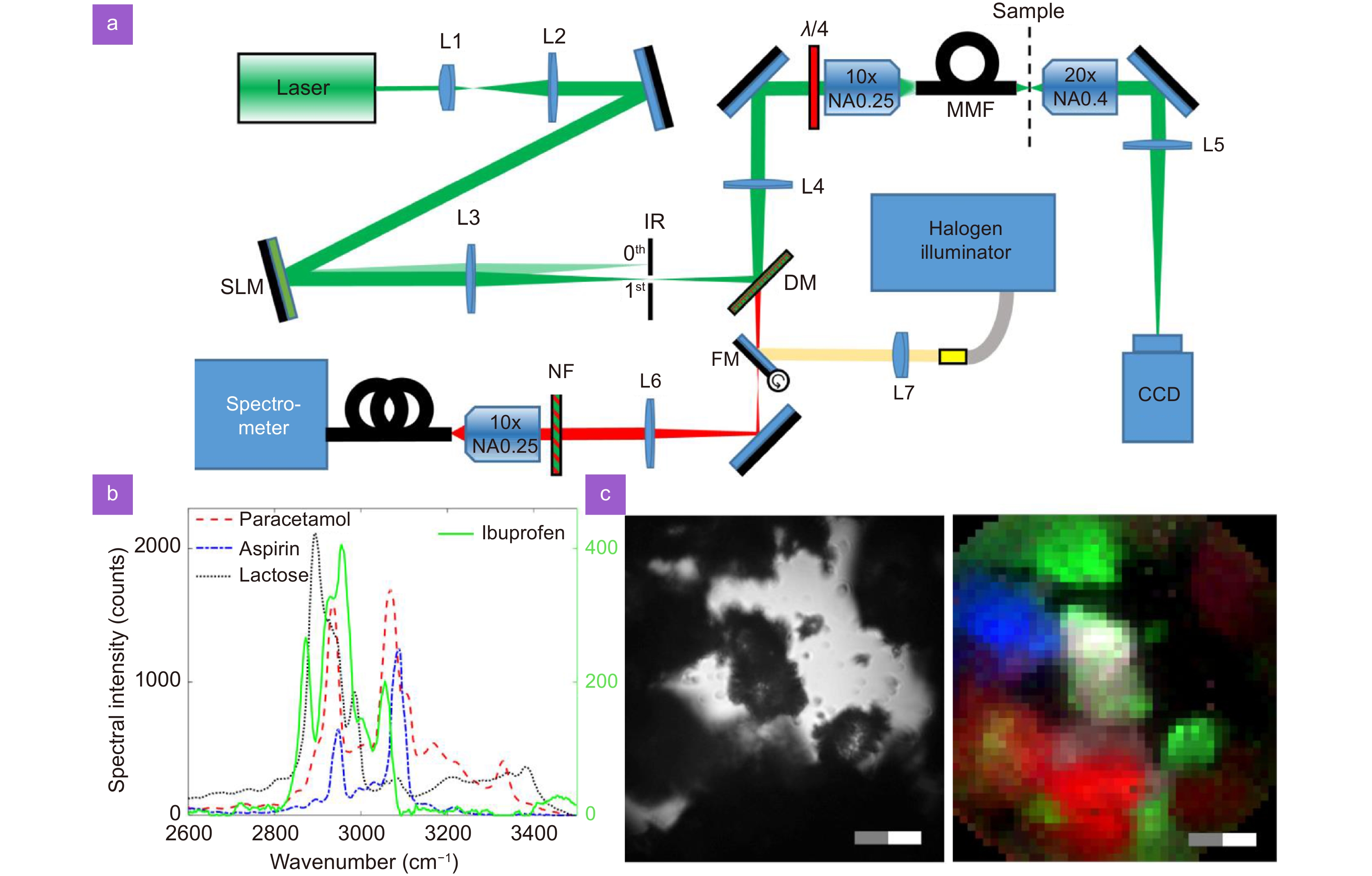
(a) Experimental setup of Raman spectroscopic imaging with the MFSE. (b) Raman spectral components of four pharmaceuticals. (c) Images of drug clusters obtained by bright field (left) and Raman spectroscopic (right) imaging modes. Figure reproduced from ref.142, under the terms of the Creative Commons Attribution 4.0 License, Optica Publication Group. Some cropping adjustments have been made to the original image.

 E-mail Alert
 RSS
RSS


 DownLoad:

