
 E-mail Alert
 RSS
RSS
| Citation: |

|
Flexible SERS substrates for hazardous materials detection: recent advances
-
Abstract
This article reviews the most recent advances in the development of flexible substrates used as surface-enhanced Raman scattering (SERS) platforms for detecting several hazardous materials (e.g., explosives, pesticides, drugs, and dyes). Different flexible platforms such as papers/filter papers, fabrics, polymer nanofibers, and cellulose fibers have been investigated over the last few years and their SERS efficacies have been evaluated. We start with an introduction of the importance of hazardous materials trace detection followed by a summary of different SERS methodologies with particular attention on flexible substrates and their advantages over the nanostructures and nanoparticle-based solid/hybrid substrates. The potential of flexible SERS substrates, in conjunction with a simple portable Raman spectrometer, is the power to enable practical/on-field/point of interest applications primarily because of their low-cost and easy sampling. -

-
References
[1] Bharati MSS. Rigid, flexible sers substrates fabricated using femtosecond laser pulses for explosives detection (Ph.D. Thesis, Submitted to University of Hyderabad, Hyderabad, India, 2020). [2] Zamora-Sequeira R, Starbird-Pérez R, Rojas-Carillo O, Vargas-Villalobos S. What are the main sensor methods for quantifying pesticides in agricultural activities? a review. Molecules 24, 2659 (2019). doi: 10.3390/molecules24142659 [3] National Research Council. Acute Exposure Guideline Levels for Selected Airborne Chemicals: Volume 3 (The National Academies Press, Washington, 2003). [4] Bartelt-Hunt SL, Knappe DRU, Barlaz MA. A review of chemical warfare agent simulants for the study of environmental behavior. Crit Rev Environ Sci Technol 38, 112–136 (2008). doi: 10.1080/10643380701643650 [5] Kumar V. Chromo-fluorogenic sensors for chemical warfare agents in real-time analysis: journey towards accurate detection and differentiation. Chem Commun 57, 3430–3444 (2021). doi: 10.1039/D1CC00132A [6] Jindal MK, Mainuddin M, Veerabuthiran S, Razdan AK. Laser based systems for standoff detection of CWA: a short review. IEEE Sens J 21, 4085–4096 (2021). doi: 10.1109/JSEN.2020.3030672 [7] Wallin S, Pettersson A, Östmark H, Hobro A. Laser-based standoff detection of explosives: a critical review. Anal Bioanal Chem 395, 259–274 (2009). doi: 10.1007/s00216-009-2844-3 [8] Mokalled L, Al-Husseini M, Kabalan KY, El-Hajj A. Sensor review for trace detection of explosives. Int J Sci Eng Res 5, 337–350 (2014). [9] Singh S. Sensors—an effective approach for the detection of explosives. J Hazard Mater 144, 15–28 (2007). doi: 10.1016/j.jhazmat.2007.02.018 [10] Ruan S, Chen YZ, Zhang P, Pan XZ, Fang C et al. Online remote monitoring of explosives by optical fibres. RSC Adv 6, 103324–103327 (2016). doi: 10.1039/C6RA24080A [11] Konstantynovski K, Njio G, Börner F, Lepcha A, Fischer T et al. Bulk detection of explosives and development of customized metal oxide semiconductor gas sensors for the identification of energetic materials. Sens Actuators B: Chem 258, 1252–1266 (2018). doi: 10.1016/j.snb.2017.11.116 [12] Wen P, Amin M, Herzog WD, Kunz RR. Key challenges and prospects for optical standoff trace detection of explosives. TrAC Trends Anal Chem 100, 136–144 (2018). doi: 10.1016/j.trac.2017.12.014 [13] Davies AG, Burnett AD, Fan WH, Linfield EH, Cunningham JE. Terahertz spectroscopy of explosives and drugs. Mater Today 11, 18–26 (2008). [14] Yinon J. Counterterrorist Detection Techniques of Explosives (Elsevier, Amsterdam, 2007). [15] Moore DS, Scharff RJ. Portable Raman explosives detection. Anal Bioanal Chem 393, 1571–1578 (2009). doi: 10.1007/s00216-008-2499-5 [16] Christesen SD, Guicheteau JA, Curtiss JM, Fountain AW. Handheld dual-wavelength Raman instrument for the detection of chemical agents and explosives. Opt Eng 55, 074103 (2016). doi: 10.1117/1.OE.55.7.074103 [17] Vandenabeele P. Practical Raman Spectroscopy: An Introduction (John Wiley & Sons, Chichester, 2013); http://doi.org/10.1002/9781119961284. [18] Mosier-Boss PA. Review of SERS substrates for chemical sensing. Nanomaterials 7, 142 (2017). doi: 10.3390/nano7060142 [19] Sun J, Gong L, Wang WJ, Gong ZJ, Wang DM et al. Surface‐enhanced Raman spectroscopy for on‐site analysis: a review of recent developments. Luminescence 35, 808–820 (2020). doi: 10.1002/bio.3796 [20] Muehlethaler C, Leona M, Lombardi JR. Review of surface enhanced Raman scattering applications in forensic science. Anal Chem 88, 152–169 (2016). doi: 10.1021/acs.analchem.5b04131 [21] Gares KL, Hufziger KT, Bykov SV, Asher SA. Review of explosive detection methodologies and the emergence of standoff deep UV resonance Raman. J Raman Spectros 47, 124–141 (2016). doi: 10.1002/jrs.4868 [22] Zhou HB, Zhang ZP, Jiang CL, Guan GJ, Zhang K et al. Trinitrotoluene explosive lights up ultrahigh Raman scattering of nonresonant molecule on a top-closed silver nanotube array. Anal Chem 83, 6913–6917 (2011). doi: 10.1021/ac201407z [23] Hakonen A, Rindzevicius T, Schmidt MS, Andersson PO, Juhlin L et al. Detection of nerve gases using surface-enhanced Raman scattering substrates with high droplet adhesion. Nanoscale 8, 1305–1308 (2016). doi: 10.1039/C5NR06524K [24] Fleischmann M, Hendra PJ, McQuillan AJ. Raman spectra of pyridine adsorbed at a silver electrode. Chem Phys Lett 26, 163–166 (1974). doi: 10.1016/0009-2614(74)85388-1 [25] Jeanmaire DL, Van Duyne RP. Surface Raman spectroelectrochemistry: part I. Heterocyclic, aromatic, and aliphatic amines adsorbed on the anodized silver electrode. J Electroanal Chem Interfacial Electrochem 84, 1–20 (1977). doi: 10.1016/S0022-0728(77)80224-6 [26] Albrecht MG, Creighton JA. Anomalously intense Raman spectra of pyridine at a silver electrode. J Am Chem Soc 99, 5215–5217 (1977). doi: 10.1021/ja00457a071 [27] Moskovits M. Surface roughness and the enhanced intensity of Raman scattering by molecules adsorbed on metals. J Chem Phys 69, 4159–4161 (1978). doi: 10.1063/1.437095 [28] Rajesh Y, Bharati MSS, Rao SV, Krishna MG. ZnO nanowire arrays decorated with titanium nitride nanoparticles as surface-enhanced Raman scattering substrates. Appl Phys A 127, 270 (2021). doi: 10.1007/s00339-021-04424-w [29] Lan LL, Gao YM, Fan XC, Li MZ, Hao Q et al. The origin of ultrasensitive SERS sensing beyond plasmonics. Front Phys 16, 43300 (2021). doi: 10.1007/s11467-021-1047-z [30] Zhen Y, Xu KC, Jiang SZ, Luo D, Chen R et al. Recent progress on two-dimensional layered materials for surface enhanced Raman spectroscopy and their applications. Mater Today Phys 18, 100378 (2021). doi: 10.1016/j.mtphys.2021.100378 [31] Basu N, Bharathi MSS, Sharma M, Yadav K, Parmar AS et al. Large area few-layer hexagonal boron nitride as a Raman enhancement material. Nanomaterials 11, 622 (2021). doi: 10.3390/nano11030622 [32] Ge MH, Pan L, Zhou GL, Chen SY, Han W et al. General surface enhanced Raman spectroscopy method for actively capturing target molecules in small gaps. J Am Chem Soc 143, 7769–7776 (2021). doi: 10.1021/jacs.1c02169 [33] Le Ru EC, Etchegoin PG. Quantifying SERS enhancements. MRS Bull 38, 631–640 (2013). doi: 10.1557/mrs.2013.158 [34] Fan MK, Andrade GFS, Brolo AG. A review on the fabrication of substrates for surface enhanced Raman spectroscopy and their applications in analytical chemistry. Anal Chim Acta 693, 7–25 (2011). doi: 10.1016/j.aca.2011.03.002 [35] Mahadeva SK, Walus K, Stoeber B. Paper as a platform for sensing applications and other devices: a review. ACS Appl Mater Interfaces 7, 8345–8362 (2015). doi: 10.1021/acsami.5b00373 [36] Restaino SM, White IM. A critical review of flexible and porous SERS sensors for analytical chemistry at the point-of-sample. Anal Chim Acta 1060, 17–29 (2019). doi: 10.1016/j.aca.2018.11.057 [37] Senthamizhan A, Balusamy B, Uyar T. Glucose sensors based on electrospun nanofibers: a review. Anal Bioanal Chem 408, 1285–1306 (2016). doi: 10.1007/s00216-015-9152-x [38] Hakonen A, Andersson PO, Schmidt MS, Rindzevicius T, Käll M. Explosive and chemical threat detection by surface-enhanced Raman scattering: a review. Anal Chim Acta 893, 1–13 (2015). doi: 10.1016/j.aca.2015.04.010 [39] Ogundare SA, van Zyl WE. A review of cellulose-based substrates for SERS: fundamentals, design principles, applications. Cellulose 26, 6489–6528 (2019). doi: 10.1007/s10570-019-02580-0 [40] Maddipatla D, Narakathu BB, Atashbar M. Recent progress in manufacturing techniques of printed and flexible sensors: a review. Biosensors 10, 199 (2020). doi: 10.3390/bios10120199 [41] Peng XY, Li D, Li YT, Xing HB, Deng W. Plasmonic tunable Ag-coated gold nanorod arrays as reusable SERS substrates for multiplexed antibiotics detection. J Mater Chem B 9, 1123–1130 (2021). doi: 10.1039/D0TB02540B [42] Xu KC, Yan HP, Tan CF, Lu YY, Li Y et al. Hedgehog inspired CuO nanowires/Cu2O composites for broadband visible‐light‐driven recyclable surface enhanced Raman scattering. Adv Opt Mater 6, 1701167 (2018). doi: 10.1002/adom.201701167 [43] Zhang DR, Pu HB, Huang LJ, Sun DW. Advances in flexible surface-enhanced Raman scattering (SERS) substrates for nondestructive food detection: fundamentals and recent applications. Trends Food Sci Technol 109, 690–701 (2021). doi: 10.1016/j.jpgs.2021.01.058 [44] Klapec DJ, Czarnopys G, Pannuto J. Interpol review of detection and characterization of explosives and explosives residues 2016-2019. Forensic Sci Int: Synergy 2, 670–700 (2020). doi: 10.1016/j.fsisyn.2020.01.020 [45] Li ZY, Huang X, Lu G. Recent developments of flexible and transparent SERS substrates. J Mater Chem C 8, 3956–3969 (2020). [46] Forbes TP, Krauss ST, Gillen G. Trace detection and chemical analysis of homemade fuel-oxidizer mixture explosives: emerging challenges and perspectives. TrAC Trends Anal Chem 131, 116023 (2020). doi: 10.1016/j.trac.2020.116023 [47] Wu JJ, Zhang L, Huang F, Ji XX, Dai HQ et al. Surface enhanced Raman scattering substrate for the detection of explosives: construction strategy and dimensional effect. J Hazard Mater 387, 121714 (2020). doi: 10.1016/j.jhazmat.2019.121714 [48] Shvalya V, Filipič G, Zavašnik J, Abdulhalim I, Cvelbar U. Surface-enhanced Raman spectroscopy for chemical and biological sensing using nanoplasmonics: the relevance of interparticle spacing and surface morphology. Appl Phys Rev 7, 031307 (2020). doi: 10.1063/5.0015246 [49] To KC, Ben-Jaber S, Parkin IP. Recent developments in the field of explosive trace detection. ACS Nano 14, 10804–10833 (2020). doi: 10.1021/acsnano.0c01579 [50] Pérez-Jiménez AI, Lyu DY, Lu ZX, Liu GK, Ren B. Surface-enhanced Raman spectroscopy: benefits, trade-offs and future developments. Chem Sci 11, 4563–4577 (2020). doi: 10.1039/D0SC00809E [51] Huang CC, Cheng CY, Lai YS. Paper-based flexible surface enhanced Raman scattering platforms and their applications to food safety. Trends Food Sci Technol 100, 349–358 (2020). doi: 10.1016/j.jpgs.2020.04.019 [52] Chen MP, Liu D, Du XY, Lo KH, Wang SP et al. 2D materials: excellent substrates for surface-enhanced Raman scattering (SERS) in chemical sensing and biosensing. TrAC Trends Anal Chem 130, 115983 (2020). doi: 10.1016/j.trac.2020.115983 [53] Xue JJ, Wu T, Dai YQ, Xia YN. Electrospinning and electrospun nanofibers: methods, materials, and applications. Chem Rev 119, 5298–5415 (2019). doi: 10.1021/acs.chemrev.8b00593 [54] Pilot R, Signorini R, Durante C, Orian L, Bhamidipati M et al. A review on surface-enhanced Raman scattering. Biosensors 9, 57 (2019). doi: 10.3390/bios9020057 [55] Lee HK, Lee YH, Koh CSL, Phan-Quang GC, Han XM et al. Designing surface-enhanced Raman scattering (SERS) platforms beyond hotspot engineering: emerging opportunities in analyte manipulations and hybrid materials. Chem Soc Rev 48, 731–756 (2019). doi: 10.1039/C7CS00786H [56] Xu KC, Zhou R, Takei K, Hong MH. Toward flexible surface-enhanced Raman scattering (SERS) sensors for point-of-care diagnostics. Adv Sci 6, 1900925 (2019). doi: 10.1002/advs.201900925 [57] Zhang S, Jia ZX, Liu TJ, Wei G, Su ZQ. Electrospinning nanoparticles-based materials interfaces for sensor applications. Sensors 19, 3977 (2019). doi: 10.3390/s19183977 [58] Liyanage T, Rael A, Shaffer S, Zaidi S, Goodpaster JV et al. Fabrication of a self-assembled and flexible SERS nanosensor for explosive detection at parts-per-quadrillion levels from fingerprints. Analyst 143, 2012–2022 (2018). doi: 10.1039/C8AN00008E [59] Xu KC, Zhang CT, Lu TH, Wang PQ, Zhou R et al. Hybrid metal-insulator-metal structures on Si nanowires array for surface enhanced Raman scattering. Opto-Electron Eng 44, 185–191 (2017). [60] Fierro-Mercado PM, Hernández-Rivera SP. Highly sensitive filter paper substrate for SERS trace explosives detection. Int J Spectrosc 2012, 716527 (2012). [61] Cui H, Li SY, Deng SZ, Chen HJ, Wang CX. Flexible, transparent, and free-standing silicon nanowire SERS platform for in situ food inspection. ACS Sens 2, 386–393 (2017). doi: 10.1021/acssensors.6b00712 [62] Jiang JL, Zou SM, Ma LW, Wang SF, Liao JS et al. Surface-enhanced Raman scattering detection of pesticide residues using transparent adhesive tapes and coated silver nanorods. ACS Appl Mater Interfaces 10, 9129–9135 (2018). doi: 10.1021/acsami.7b18039 [63] Lee M, Oh K, Choi HK, Lee SG, Youn HJ et al. Subnanomolar sensitivity of filter paper-based SERS sensor for pesticide detection by hydrophobicity change of paper surface. ACS Sens 3, 151–159 (2018). doi: 10.1021/acssensors.7b00782 [64] Wang KQ, Sun DW, Pu HB, Wei QY, Huang LJ. Stable, flexible, and high-performance SERS chip enabled by a ternary film-packaged plasmonic nanoparticle array. ACS Appl Mater Interfaces 11, 29177–29186 (2019). doi: 10.1021/acsami.9b09746 [65] Lin Y, Gritsenko D, Liu Q, Lu XN, Xu J. Recent advancements in functionalized paper-based electronics. ACS Appl Mater Interfaces 8, 20501–20515 (2016). doi: 10.1021/acsami.6b04854 [66] Kumar A, Santhanam V. Paper swab based SERS detection of non-permitted colourants from dals and vegetables using a portable spectrometer. Anal Chim Acta 1090, 106–113 (2019). doi: 10.1016/j.aca.2019.08.073 [67] Liu Q, Wang JH, Wang BK, Li Z, Huang H et al. Paper-based plasmonic platform for sensitive, noninvasive, and rapid cancer screening. Biosens Bioelectron 54, 128–134 (2014). doi: 10.1016/j.bios.2013.10.067 [68] Kim EJ, Kim H, Park E, Kim T, Chung DR et al. Paper-based multiplex surface-enhanced Raman scattering detection using polymerase chain reaction probe codification. Anal Chem 93, 3677–3685 (2021). doi: 10.1021/acs.analchem.0c05285 [69] Park M, Jung H, Jeong Y, Jeong KH. Plasmonic schirmer strip for human tear-based gouty arthritis diagnosis using surface-enhanced Raman scattering. ACS Nano 11, 438–443 (2017). doi: 10.1021/acsnano.6b06196 [70] Gao RK, Song XF, Zhan CB, Weng CG, Cheng S et al. Light trapping induced flexible wrinkled nanocone SERS substrate for highly sensitive explosive detection. Sens Actuators B: Chem 314, 128081 (2020). doi: 10.1016/j.snb.2020.128081 [71] Lee CH, Tian LM, Singamaneni S. Paper-based SERS swab for rapid trace detection on real-world surfaces. ACS Appl Mater Interfaces 2, 3429–3435 (2010). doi: 10.1021/am1009875 [72] Yu WW, White IM. Chromatographic separation and detection of target analytes from complex samples using inkjet printed SERS substrates. Analyst 138, 3679–3686 (2013). doi: 10.1039/c3an00673e [73] Polavarapu L, Porta AL, Novikov SM, Coronado-Puchau M, Liz-Marzán ML. Pen on paper approach toward the design of universal surface enhanced Raman scattering substrates. Small 10, 3065–3071 (2014). doi: 10.1002/smll.201400438 [74] Lan LL, Hou XY, Gao YM, Fan XC, Qiu T. Inkjet-printed paper-based semiconducting substrates for surface-enhanced Raman spectroscopy. Nanotechnology 31, 055502 (2020). doi: 10.1088/1361-6528/ab4f11 [75] Chamuah N, Hazarika A, Hatiboruah D, Nath P. SERS on paper: an extremely low cost technique to measure Raman signal. J Phys D: Appl Phys 50, 485601 (2017). doi: 10.1088/1361-6463/aa8fef [76] Yu CC, Chou SY, Tseng YC, Tseng SC, Yen YT et al. Single-shot laser treatment provides quasi-three-dimensional paper-based substrates for SERS with attomolar sensitivity. Nanoscale 7, 1667–1677 (2015). doi: 10.1039/C4NR05178E [77] Zhang R, Xu BB, Liu XQ, Zhang YL, Xu Y et al. Highly efficient SERS test strips. Chem Commun 48, 5913–5915 (2012). doi: 10.1039/c2cc31604h [78] Raza A, Saha B. In situ silver nanoparticles synthesis in agarose film supported on filter paper and its application as highly efficient SERS test stripes. Forensic Sci Int 237, e42–e46 (2014). doi: 10.1016/j.forsciint.2014.01.019 [79] Das D, Senapati S, Nanda KK. “Rinse, Repeat”: an efficient and reusable SERS and catalytic platform fabricated by controlled deposition of silver nanoparticles on cellulose paper. ACS Sustainable Chem Eng 7, 14089–14101 (2019). doi: 10.1021/acssuschemeng.9b02651 [80] Oliveira MJ, Quaresma P, de Almeida MP, Araújo A, Pereira E et al. Office paper decorated with silver nanostars - an alternative cost effective platform for trace analyte detection by SERS. Sci Rep 7, 2480 (2017). doi: 10.1038/s41598-017-02484-8 [81] Yu WW, White IM. Inkjet printed surface enhanced Raman spectroscopy array on cellulose paper. Anal Chem 82, 9626–9630 (2010). doi: 10.1021/ac102475k [82] Li YX, Zhang K, Zhao JJ, Ji J, Ji C et al. A three dimensional silver nanoparticles decorated plasmonic paper strip for SERS detection of low-abundance molecules. Talanta 147, 493–500 (2016). doi: 10.1016/j.talanta.2015.10.025 [83] Marques PAAP, Nogueira HIS, Pinto RJB, Neto CP, Trindade T. Silver‐bacterial cellulosic sponges as active SERS substrates. J Raman Spectrosc 39, 439–443 (2008). doi: 10.1002/jrs.1853 [84] Kim W, Kim YH, Park HK, Choi S. Facile fabrication of a silver nanoparticle immersed, surface-enhanced Raman scattering imposed paper platform through successive ionic layer absorption and reaction for on-site bioassays. ACS Appl Mater Interfaces 7, 27910–27917 (2015). doi: 10.1021/acsami.5b09982 [85] Hasi WLJ, Lin X, Lou XT, Lin S, Yang F et al. Chloride ion assisted self assembly of silver nanoparticles on filter paper as SERS substrate. Appl Phys A 118, 799–807 (2015). doi: 10.1007/s00339-014-8800-x [86] Huang ZF, Cao G, Sun Y, Du SR, Li YZ et al. Evaluation and optimization of paper-based SERS substrate for potential label-free Raman analysis of seminal plasma. J Nanomater 2017, 4807064 (2017). [87] Mehn D, Morasso C, Vanna R, Bedoni M, Prosperi D et al. Immobilised gold nanostars in a paper-based test system for surface-enhanced Raman spectroscopy. Vib Spectrosc 68, 45–50 (2013). doi: 10.1016/j.vibspec.2013.05.010 [88] Wang C, Liu BX, Dou XC. Silver nanotriangles-loaded filter paper for ultrasensitive SERS detection application benefited by interspacing of sharp edges. Sens Actuators B: Chem 231, 357–364 (2016). doi: 10.1016/j.snb.2016.03.030 [89] Lee CH, Hankus ME, Tian LM, Pellegrino PM, Singamaneni S. Highly sensitive surface enhanced Raman scattering substrates based on filter paper loaded with plasmonic nanostructures. Anal Chem 83, 8953–8958 (2011). doi: 10.1021/ac2016882 [90] Hoppmann EP, Yu WW, White IM. Highly sensitive and flexible inkjet printed SERS sensors on paper. Methods 63, 219–224 (2013). doi: 10.1016/j.ymeth.2013.07.010 [91] Hoppmann EP, Yu WW, White IM. Inkjet-printed fluidic paper devices for chemical and biological analytics using surface enhanced Raman spectroscopy. IEEE J Sel Top Quantum Electron 20, 7300510 (2014). [92] Kim D, Gwon G, Lee G, Jeon Y, Kim UJ et al. Surface enhanced Raman scattering active AuNR array cellulose films for multi hazard detection. J Hazard Mater 402, 123505 (2021). doi: 10.1016/j.jhazmat.2020.123505 [93] Xian L, You RY, Lu DC, Wu CJ, Feng SY et al. Surface-modified paper-based SERS substrates for direct-droplet quantitative determination of trace substances. Cellulose 27, 1483–1495 (2020). doi: 10.1007/s10570-019-02855-6 [94] Moram SSB, Byram C, Shibu SN, Chilukamarri BM, Soma VR. Ag/Au nanoparticle-loaded paper-based versatile surface-enhanced Raman spectroscopy substrates for multiple explosives detection. ACS Omega 3, 8190–8201 (2018). doi: 10.1021/acsomega.8b01318 [95] Lin S, Hasi W, Han SQGW, Lin X, Wang L. A dual-functional PDMS-assisted paper-based SERS platform for the reliable detection of thiram residue both on fruit surfaces and in juice. Anal Methods 12, 2571–2579 (2020). doi: 10.1039/D0AY00483A [96] Ming HZ, Zhang YZ, Kotaki M, Ramakrishna S. A review on polymer nanofibers by electrospinning and their applications in nanocomposites. Compos Sci Technol 63, 2223–2253 (2003). doi: 10.1016/S0266-3538(03)00178-7 [97] Teo WE, Ramakrishna S. A review on electrospinning design and nanofibre assemblies. Nanotechnology 17, R89–R106 (2006). doi: 10.1088/0957-4484/17/14/R01 [98] Aleisa R. Electrospinning. In Handbook of Synthetic Methodologies and Protocols of Nanomaterials, Liu YD, He L, Yin YD edn, 149–181 (World Scientific, 2019); http://doi.org/10.1142/9789813277847_0006. [99] Husain O, Lau W, Edirisinghe M, Parhizkar M. Investigating the particle to fibre transition threshold during electrohydrodynamic atomization of a polymer solution. Mater Sci Eng: C 65, 240–250 (2016). doi: 10.1016/j.msec.2016.03.076 [100] Wan MH, Zhao HD, Peng LC, Zou XY, Zhao YB et al. Loading of Au/Ag bimetallic nanoparticles within and outside of the flexible SiO2 electrospun nanofibers as highly sensitive, stable, repeatable substrates for versatile and trace SERS detection. Polymers 12, 3008 (2020). doi: 10.3390/polym12123008 [101] Zhang ZJ, Wu YP, Wang ZH, Zou XY, Zhao YB et al. Fabrication of silver nanoparticles embedded into polyvinyl alcohol (Ag/PVA) composite nanofibrous films through electrospinning for antibacterial and surface-enhanced Raman scattering (SERS) activities. Mater Sci Eng: C 69, 462–469 (2016). doi: 10.1016/j.msec.2016.07.015 [102] Jalaja K, Bhuvaneswari S, Ganiga M, Divyamol R, Anup S et al. Effective SERS detection using a flexible wiping substrate based on electrospun polystyrene nanofibers. Anal Methods 9, 3998–4003 (2017). doi: 10.1039/C7AY00882A [103] Jia P, Chang J, Wang JQ, Zhang P, Cao B et al. Fabrication and formation mechanism of Ag nanoplate‐decorated nanofiber mats and their application in SERS. Chem-Asian J 11, 86–92 (2016). doi: 10.1002/asia.201500777 [104] Chamuah N, Bhuyan N, Das PP, Ojah N, Choudhary AJ et al. Gold-coated electrospun PVA nanofibers as SERS substrate for detection of pesticides. Sens Actuators B: Chem 273, 710–717 (2018). doi: 10.1016/j.snb.2018.06.079 [105] Motamedi AS, Mirzadeh H, Hajiesmaeilbaigi F, Bagheri-Khoulenjani S, Shokrgozar MA. Piezoelectric electrospun nanocomposite comprising Au NPs/PVDF for nerve tissue engineering. J Biomed Mater Res Part A 105, 1984–1993 (2017). doi: 10.1002/jbm.a.36050 [106] Zhang CL, Lv KP, Cong HP, Yu SH. Controlled assemblies of gold nanorods in PVA nanofiber matrix as flexible free‐standing SERS substrates by electrospinning. Small 8, 648–653 (2012). doi: 10.1002/smll.201102230 [107] Zhang LF, Gong X, Bao Y, Zhao Y, Xi M et al. Electrospun nanofibrous membranes surface-decorated with silver nanoparticles as flexible and active/sensitive substrates for surface-enhanced Raman scattering. Langmuir 28, 14433–14440 (2012). doi: 10.1021/la302779q [108] Alyami A, Quinn AJ, Iacopino D. Flexible and transparent Surface Enhanced Raman Scattering (SERS)-Active Ag NPs/PDMS composites for in-situ detection of food contaminants. Talanta 201, 58–64 (2019). doi: 10.1016/j.talanta.2019.03.115 [109] Qiu HW, Wang MQ, Jiang SZ, Zhang L, Yang Z et al. Reliable molecular trace-detection based on flexible SERS substrate of graphene/Ag-nanoflowers/PMMA. Sens Actuators B: Chem 249, 439–450 (2017). doi: 10.1016/j.snb.2017.04.118 [110] Zuo ZW, Zhu K, Gu C, Wen YB, Cui GL et al. Transparent, flexible surface enhanced Raman scattering substrates based on Ag coated structured PET (polyethylene terephthalate) for in-situ detection. Appl Surf Sci 379, 66–72 (2016). doi: 10.1016/j.apsusc.2016.04.022 [111] Creedon NC, Lovera P, Furey A, O’Riordan A. Transparent polymer-based SERS substrates templated by a soda can. Sens Actuators B: Chem 259, 64–74 (2018). doi: 10.1016/j.snb.2017.12.039 [112] Alvarez-Ruiz DT, Almohammed S, Fularz A, Barwich ST, Rice JH. Self-energized organic-inorganic hybrid composite for surface enhanced Raman spectroscopy. J Appl Phys 129, 193102 (2021). doi: 10.1063/5.0048802 [113] Wang KQ, Sun DW, Pu HB, Wei QY. Polymer multilayers enabled stable and flexible Au@Ag nanoparticle array for nondestructive SERS detection of pesticide residues. Talanta 223, 121782 (2021). doi: 10.1016/j.talanta.2020.121782 [114] Zhang CP, Yi PY, Peng LF, Lai XM, Chen J et al. Continuous fabrication of nanostructure arrays for flexible surface enhanced Raman scattering substrate. Sci Rep 7, 39814 (2017). doi: 10.1038/srep39814 [115] Fang LN, Li JC, Zhang JR, Han DD. Femtosecond laser structuring for flexible surface-enhanced Raman spectroscopy substrates. IEEE Photonics J 13, 6800908 (2021). [116] Byram C, Moram SSB, Soma VR. SERS based detection of multiple analytes from dye/explosive mixtures using picosecond laser fabricated gold nanoparticles and nanostructures. Analyst 144, 2327–2336 (2019). doi: 10.1039/C8AN01276H [117] Naqvi TK, Bajpai A, Bharati MSS, Kulkarni MM, Siddiqui AM et al. Ultra-sensitive reusable SERS sensor for multiple hazardous materials detection on single platform. J Hazard Mater 407, 124353 (2021). doi: 10.1016/j.jhazmat.2020.124353 [118] Byram C, Moram SSB, Soma VR. Picosecond laser fabricated Ag, Au and Ag-Au nanoparticles for detecting ammonium perchlorate using a portable Raman spectrometer. AIP Conf Proc 1942, 050028 (2018). [119] Hamad S, Moram SSB, Yendeti B, Podagatlapalli GK, Rao SVSN et al. Femtosecond laser-induced, nanoparticle-embedded periodic surface structures on crystalline silicon for reproducible and multi-utility SERS platforms. ACS Omega 3, 18420–18432 (2018). doi: 10.1021/acsomega.8b02629 [120] Moram SSB, Shaik AK, Byram C, Hamad S, Soma VR. Instantaneous trace detection of nitro-explosives and mixtures with nanotextured silicon decorated with Ag–Au alloy nanoparticles using the SERS technique. Anal Chim Acta 1101, 157–168 (2020). doi: 10.1016/j.aca.2019.12.026 [121] Podagatlapalli GK, Hamad S, Mohiddon MA, Rao SV. Effect of oblique incidence on silver nanomaterials fabricated in water via ultrafast laser ablation for photonics and explosives detection. Appl Surf Sci 303, 217–232 (2014). doi: 10.1016/j.apsusc.2014.02.152 [122] Tang B, Wang JF, Xu SP, Afrin T, Xu WQ et al. Application of anisotropic silver nanoparticles: multifunctionalization of wool fabric. J Colloid Interface Sci 356, 513–518 (2011). doi: 10.1016/j.jcis.2011.01.054 [123] Liu J, Zhou J, Tang B, Zeng T, Li YL et al. Surface enhanced Raman scattering (SERS) fabrics for trace analysis. Appl Surf Sci 386, 296–302 (2016). doi: 10.1016/j.apsusc.2016.05.150 [124] Chen YM, Ge FY, Guang SY, Cai ZS. Low-cost and large-scale flexible SERS-cotton fabric as a wipe substrate for surface trace analysis. Appl Surf Sci 436, 111–116 (2018). doi: 10.1016/j.apsusc.2017.11.288 [125] Zheng YD, Xiao MD, Jiang SX, Ding F, Wang JF. Coating fabrics with gold nanorods for colouring, UV-protection, and antibacterial functions. Nanoscale 5, 788–795 (2013). doi: 10.1039/C2NR33064D [126] Wang RH, Wang XW, Xin JH. Advanced visible-light-driven self-cleaning cotton by Au/TiO2/SiO2 photocatalysts. ACS Appl Mater Interfaces 2, 82–85 (2010). doi: 10.1021/am900588s [127] Gao W, Xu JT, Cheng C, Qiu S, Jiang SX. Rapid and highly sensitive SERS detection of fungicide based on flexible “wash free” metallic textile. Appl Surf Sci 512, 144693 (2020). doi: 10.1016/j.apsusc.2019.144693 [128] Lu SC, You TT, Yang N, Gao YK, Yin PG. Flexible SERS substrate based on Ag nanodendrite–coated carbon fiber cloth: simultaneous detection for multiple pesticides in liquid droplet. Anal Bioanal Chem 412, 1159–1167 (2020). doi: 10.1007/s00216-019-02344-6 [129] Zhang ZL, Si TT, Liu J, Zhou GW. In-situ grown silver nanoparticles on nonwoven fabrics based on mussel-inspired polydopamine for highly sensitive SERS Carbaryl pesticides detection. Nanomaterials 9, 384 (2019). doi: 10.3390/nano9030384 [130] Liu Y, Zhang Y, Tardivel M, Lequeux M, Chen XP et al. Evaluation of the reliability of six commercial SERS substrates. Plasmonics 15, 743–752 (2020). doi: 10.1007/s11468-019-01084-8 [131] Hakonen A, Wu KY, Schmidt MS, Andersson PO, Boisen A et al. Detecting forensic substances using commercially available SERS substrates and handheld Raman spectrometers. Talanta 189, 649–652 (2018). doi: 10.1016/j.talanta.2018.07.009 [132] Wang JP, Yang L, Liu BH, Jiang HH, Liu RY et al. Inkjet-printed silver nanoparticle paper detects airborne species from crystalline explosives and their ultratrace residues in open environment. Anal Chem 86, 3338–3345 (2014). doi: 10.1021/ac403409q [133] Moram SSB, Byram C, Soma VR. Gold-nanoparticle-and nanostar-loaded paper-based SERS substrates for sensing nanogram-level Picric acid with a portable Raman spectrometer. Bull Mater Sci 43, 53 (2020). doi: 10.1007/s12034-019-2017-8 [134] Khan GA, Demirtaş Ö, Demir AK, Ayteki Ö, Bek A et al. Fabrication of flexible, cost-effective, and scalable silver substrates for efficient surface enhanced Raman spectroscopy based trace detection. Colloids Surf A: Physicochem Eng Aspects 619, 126542 (2021). doi: 10.1016/j.colsurfa.2021.126542 [135] Yu WW, White IM. Inkjet-printed paper-based SERS dipsticks and swabs for trace chemical detection. Analyst 138, 1020–1025 (2013). doi: 10.1039/C2AN36116G [136] Alder R, Hong J, Chow E, Fang JH, Isa F et al. Application of plasma-printed paper-based SERS substrate for cocaine detection. Sensors 21, 810 (2021). doi: 10.3390/s21030810 [137] Zhang LZ, Liu J, Zhou GW, Zhang ZL. Controllable in-situ growth of silver nanoparticles on filter paper for flexible and highly sensitive SERS sensors for malachite green residue detection. Nanomaterials 10, 826 (2020). doi: 10.3390/nano10050826 [138] Lee CW, Chia ZC, Hsieh YT, Tsai HC, Tai Y et al. A facile wet-chemistry approach to engineer an Au-based SERS substrate and enhance sensitivity down to ppb-level detection. Nanoscale 13, 3991–3999 (2021). doi: 10.1039/D0NR06537D [139] Zhu YQ, Li MQ, Yu DY, Yang LB. A novel paper rag as ‘D-SERS’substrate for detection of pesticide residues at various peels. Talanta 128, 117–124 (2014). doi: 10.1016/j.talanta.2014.04.066 [140] Bolz A, Panne U, Rurack K, Buurman M. Glass fibre paper-based test strips for sensitive SERS sensing. Anal Methods 8, 1313–1318 (2016). doi: 10.1039/C5AY03096J [141] Zhu JJ, Chen QS, Kutsanedzie FYH, Yang MX, Ouyang Q et al. Highly sensitive and label-free determination of thiram residue using surface-enhanced Raman spectroscopy (SERS) coupled with paper-based microfluidics. Anal Methods 9, 6186–6193 (2017). doi: 10.1039/C7AY01637A [142] Ma YD, Wang YH, Luo Y, Duan HZ, Li D et al. Rapid and sensitive on-site detection of pesticide residues in fruits and vegetables using screen-printed paper-based SERS swabs. Anal Methods 10, 4655–4664 (2018). doi: 10.1039/C8AY01698D [143] Sun J, Gong L, Lu YT, Wang DM, Gong ZJ et al. Dual functional PDMS sponge SERS substrate for the on-site detection of pesticides both on fruit surfaces and in juice. Analyst 143, 2689–2695 (2018). doi: 10.1039/C8AN00476E [144] Kim D, Ko Y, Kwon G, Choo YM, You J. Low-cost, high-performance plasmonic nanocomposites for hazardous chemical detection using surface enhanced Raman scattering. Sens Actuators B: Chem 274, 30–36 (2018). doi: 10.1016/j.snb.2018.07.107 [145] Luo W, Chen M, Hao NY, Huang XQ, Zhao XY et al. In situ synthesis of gold nanoparticles on pseudo-paper films as flexible SERS substrate for sensitive detection of surface organic residues. Talanta 197, 225–233 (2019). doi: 10.1016/j.talanta.2018.12.099 [146] Atanasov PA, Nedyalkov NN, Fukata N, Jevasuwan W, Subramani T et al. Surface-enhanced Raman spectroscopy (SERS) of mancozeb and thiamethoxam assisted by gold and silver nanostructures produced by laser techniques on paper. Appl Spectros 73, 313–319 (2019). doi: 10.1177/0003702818816304 [147] Zhang CM, You TT, Yang N, Gao YK, Jiang L et al. Hydrophobic paper-based SERS platform for direct-droplet quantitative determination of melamine. Food Chem 287, 363–368 (2019). doi: 10.1016/j.foodchem.2019.02.094 [148] Xie J, Li LY, Khan IM, Wang ZP, Ma XY. Flexible paper-based SERS substrate strategy for rapid detection of methyl parathion on the surface of fruit. Spectrochim Acta Part A: Mol Biomol Spectrosc 231, 118104 (2020). doi: 10.1016/j.saa.2020.118104 [149] Chen J, Huang MZ, Kong LL. Flexible Ag/nanocellulose fibers SERS substrate and its applications for in-situ hazardous residues detection on food. Appl Surf Sci 533, 147454 (2020). doi: 10.1016/j.apsusc.2020.147454 [150] Sun L, Yu ZL, Alsammarraie FK, Lin MH, Kong FB et al. Development of cellulose nanofiber-based substrates for rapid detection of ferbam in kale by surface-enhanced Raman spectroscopy. Food Chem 347, 129023 (2021). doi: 10.1016/j.foodchem.2021.129023 [151] Song SW, Kim D, Kim J, You J, Kim HM. Flexible nanocellulose-based SERS substrates for fast analysis of hazardous materials by spiral scanning. J Hazard Mater 414, 125160 (2021). doi: 10.1016/j.jhazmat.2021.125160 [152] Tay LL, Poirier S, Ghaemi A, Hulse J, Wang SL. Paper-based surface-enhanced Raman spectroscopy sensors for field applications. J Raman Spectros 52, 563–572 (2021). doi: 10.1002/jrs.6017 [153] Lee HG, Choi W, Yang SY, Kim DH, Park SG et al. PCR-coupled Paper-based surface-enhanced Raman scattering (SERS) sensor for rapid and sensitive detection of respiratory bacterial DNA. Sens Actuators B: Chem 326, 128802 (2021). doi: 10.1016/j.snb.2020.128802 [154] Kim D, Kim J, Henzie J, Ko Y, Lim H et al. Mesoporous Au films assembled on flexible cellulose nanopaper as high-performance SERS substrates. Chem Eng J 419, 129445 (2021). doi: 10.1016/j.cej.2021.129445 [155] Xu XY, Hu XM, Fu FY, Liu L, Liu XD. DNA-induced assembly of silver nanoparticle decorated cellulose nanofiber: a flexible surface-enhanced Raman spectroscopy substrate for the selective charge molecular detection and wipe test of pesticide residues in fruits. ACS Sustainable Chem Eng 9, 5217–5229 (2021). doi: 10.1021/acssuschemeng.1c00788 [156] Gong ZJ, Du HJ, Cheng FS, Wang C, Wang CC et al. Fabrication of SERS swab for direct detection of trace explosives in fingerprints. ACS Appl Mater Interfaces 6, 21931–21937 (2014). doi: 10.1021/am507424v [157] Wang C, Wong KW, Wang Q, Zhou YF, Tang CY et al. Silver-nanoparticles-loaded chitosan foam as a flexible SERS substrate for active collecting analytes from both solid surface and solution. Talanta 191, 241–247 (2019). doi: 10.1016/j.talanta.2018.08.067 [158] Kong LL, Huang MZ, Chen J, Lin MS. Fabrication of sensitive silver-decorated cotton swabs for SERS quantitative detection of mixed pesticide residues in bitter gourds. New J Chem 44, 12779–12784 (2020). doi: 10.1039/D0NJ02054K [159] Liu J, Si TT, Zhang ZL. Mussel-inspired immobilization of silver nanoparticles toward sponge for rapid swabbing extraction and SERS detection of trace inorganic explosives. Talanta 204, 189–197 (2019). doi: 10.1016/j.talanta.2019.05.110 [160] Bharati MSS, Byram C, Banerjee D, Sarma D, Barkakaty B et al. Gold nanoparticle nanofibres as SERS substrate for detection of methylene blue and a chemical warfare simulant (methyl salicylate). Bull Mater Sci 14, 103 (2021). [161] Chen Y, Cao JL, Wei HY, Wu ZG, Wang XP et al. Synthesis of polyvinyl alcohol/Ag electrospun nanofibers as highly efficient flexible SERS substrates. Vib Spectrosc 114, 103246 (2021). doi: 10.1016/j.vibspec.2021.103246 [162] Kong LS, Dong NX, Tian GF, Qi SL, Wu DZ. Highly enhanced Raman scattering with good reproducibility observed on a flexible PI nanofabric substrate decorated by silver nanoparticles with controlled size. Appl Surf Sci 511, 145443 (2020). doi: 10.1016/j.apsusc.2020.145443 [163] Cai LM, Deng Z, Dong J, Song SD, Wang YR et al. Fabrication of non-woven fabric-based SERS substrate for direct detection of pesticide residues in fruits. J Anal Test 1, 322–329 (2017). doi: 10.1007/s41664-017-0035-3 [164] Cheng DS, He MT, Ran JH, Cai GM, Wu JH et al. Depositing a flexible substrate of triangular silver nanoplates onto cotton fabrics for sensitive SERS detection. Sens Actuators B: Chem 270, 508–517 (2018). doi: 10.1016/j.snb.2018.05.075 [165] Cheng DS, Bai X, He MT, Wu JH, Yang HJ et al. Polydopamine-assisted immobilization of Ag@AuNPs on cotton fabrics for sensitive and responsive SERS detection. Cellulose 26, 4191–4204 (2019). doi: 10.1007/s10570-019-02343-x [166] Bian XY, Xu JT, Yang J, Chiu KI, Jiang SX. Flexible Ag SERS substrate for non-destructive and rapid detection of toxic materials on irregular surface. Surf Interfaces 23, 100995 (2021). doi: 10.1016/j.surfin.2021.100995 [167] Zheng WW, Tian WT, Liu XJ, Zhang QQ, Zong CH et al. In situ photochemical deposition of Ag nanoparticles on polyester fiber membranes as flexible SERS substrates for sensitive detection of sodium saccharin in soft drinks. Microchem J 164, 106003 (2021). doi: 10.1016/j.microc.2021.106003 [168] Tian XR, Zhai P, Guo JQ, Yu Q, Xu LZ et al. Fabrication of plasmonic cotton gauze-Ag composite as versatile SERS substrate for detection of pesticides residue. Spectrochim Acta Part A: Mol Biomol Spectrosc 257, 119766 (2021). doi: 10.1016/j.saa.2021.119766 [169] Liu AR, Zhang S, Guang SY, Ge FY, Wang J. Ag-coated nylon fabrics as flexible substrates for surface-enhanced Raman scattering swabbing applications. J Mater Res 35, 1271–1278 (2020). doi: 10.1557/jmr.2020.103 [170] Ning S, Wang ZK, Mu J, Jie Z. Flexible carbon fiber cloth decorated by Ag nanoparticles for high Raman enhancement. Opt Mater Express 11, 1321–1333 (2021). [171] Sun MM, Qian HM, Liu J, Li YC, Pang SP et al. A flexible conductive film prepared by the oriented stacking of Ag and Au/Ag alloy nanoplates and its chemically roughened surface for explosive SERS detection and cell adhesion. RSC Adv 7, 7073–7078 (2017). doi: 10.1039/C6RA25956A [172] Gupta P, Luan JY, Wang ZY, Cao SS, Bae SH et al. On-demand electromagnetic hotspot generation in surface-enhanced Raman scattering substrates via “add-on” plasmonic patch. ACS Appl Mater Interfaces 11, 37939–37946 (2019). doi: 10.1021/acsami.9b12402 [173] Emamian S, Eshkeiti A, Narakathu BB, Avuthu SGR, Atashbar MZ. Gravure printed flexible surface enhanced Raman spectroscopy (SERS) substrate for detection of 2,4-dinitrotoluene (DNT) vapor. Sens Actuators B: Chem 217, 129–135 (2015). doi: 10.1016/j.snb.2014.10.069 [174] Liu W, Song ZH, Zhao YF, Liu Y, He X et al. Flexible porous aerogels decorated with Ag nanoparticles as an effective SERS substrate for label-free trace explosives detection. Anal Methods 12, 4123–4129 (2020). doi: 10.1039/D0AY00771D [175] Gao RK, Qian HY, Weng CG, Wang XL, Xie C et al. A SERS stamp: multiscale coupling effect of silver nanoparticles and highly ordered nano-micro hierarchical substrates for ultrasensitive explosive detection. Sens Actuators B: Chem 321, 128543 (2020). doi: 10.1016/j.snb.2020.128543 [176] Xu KC, Wang ZY, Tan CF, Kang N, Chen LW et al. Uniaxially stretched flexible surface plasmon resonance film for versatile surface enhanced Raman scattering diagnostics. ACS Appl Mater Interfaces 9, 26341–26349 (2017). doi: 10.1021/acsami.7b06669 [177] Li CH, Xu SC, Yu J, Jiang SZ, Liu AH et al. 3D hybrid MoS2/AgNPs/inverted pyramid PMMA resonant cavity system for the excellent flexible surface enhanced Raman scattering sensor. Sens Actuators B: Chem 274, 152–162 (2018). doi: 10.1016/j.snb.2018.07.150 [178] Xiu XW, Guo Y, Li CH, Li Z, Li DZ et al. High-performance 3D flexible SERS substrate based on graphene oxide/silver nanoparticles/pyramid PMMA. Opt Mater Express 8, 844–857 (2018). doi: 10.1364/OME.8.000844 [179] Zang SY, Liu H, Wang Q, Yang JW, Pang ZQ et al. Facile fabrication of Au nanoworms covered polyethylene terephthalate (PET) film: towards flexible SERS substrates. Mater Lett 294, 129643 (2021). doi: 10.1016/j.matlet.2021.129643 [180] Li Y, Xin XL, Zhang TT, Li WH, Li JS et al. Raspberry like polyamide@ Ag hybrid nanoarrays with flexible cores and SERS signal enhancement strategy for adenosine detection. Chem Eng J 422, 129983 (2021). doi: 10.1016/j.cej.2021.129983 [181] Chen JM, Huang YJ, Kannan P, Zhang L, Lin ZY et al. Flexible and adhesive surface enhance Raman scattering active tape for rapid detection of pesticide residues in fruits and vegetables. Anal Chem 88, 2149–2155 (2016). doi: 10.1021/acs.analchem.5b03735 [182] Wei W, Du YX, Zhang LM, Yang Y, Gao YF. Improving SERS hot spots for on-site pesticide detection by combining silver nanoparticles with nanowires. J Mater Chem C 6, 8793–8803 (2018). [183] Ma Y, Chen Y, Tian YR, Gu CJ, Jiang T. Contrastive study of in situ sensing and swabbing detection based on SERS-active gold nanobush–PDMS hybrid film. J Agric Food Chem 69, 1975–1983 (2021). doi: 10.1021/acs.jafc.0c06562 [184] Xu LM, Liu HG, Hui Z, Hong MH. One-step fabrication of metal nanoparticles on polymer film by femtosecond LIPAA method for SERS detection. Talanta 228, 122204 (2021). doi: 10.1016/j.talanta.2021.122204 [185] Sitjar J, Liao JD, Lee H, Pan LP, Liu BH et al. Ag nanostructures with spikes on adhesive tape as a flexible sers-active substrate for in situ trace detection of pesticides on fruit skin. Nanomaterials 9, 1750 (2019). doi: 10.3390/nano9121750 [186] https://www.stellarnet.us/spectrometers-accessories/sers-substrates/. [187] https://www.horiba.com/en_en/products/detail/action/show/Product/sers-substrates-1635/. [188] https://www.sersitive.eu/. [189] http://enspectr.com/applications/sers-analysis/. [190] https://www.silmeco.com/products/sers-substrate-serstrate/. [191] https://www.hamamatsu.com/jp/en/product/optical-components/sers-substrate/index.html. [192] https://integratedoptics.com/products/sers-substrates. [193] https://www.trademed.com/products/6451/SERS-Substrates.html. [194] http://www.madatec.com/RAMAN_files/Q-SERS%20G1_data%20sheet-Madatec.pdf. [195] https://www.metrohm.com/en/products/607506170. [196] Langer J, de Aberasturi DJ, Aizpurua J, Alvarez-Puebla RA, Auguié B et al. Present and future of surface-enhanced Raman scattering. ACS Nano 14, 28–117 (2020). doi: 10.1021/acsnano.9b04224 [197] Goodacre R, Graham D, Faulds, K. Recent developments in quantitative SERS: Moving towards absolute quantification. Trends Anal Chem 102, 359–368 (2018). doi: 10.1016/j.trac.2018.03.005 [198] Szaniawska A, Kudelski A. Applications of Surface-Enhanced Raman Scattering in Biochemical and Medical Analysis. Front Chem 9, 664134 (2021). doi: 10.3389/fchem.2021.664134 [199] Cupil-Garcia V, Strobbia P, Crawford BM, Wang HN, Ngo H et al. Plasmonic nanoplatforms: from surface‐enhanced Raman scattering sensing to biomedical applications. J Raman Spectros 52, 541–553 (2021). doi: 10.1002/jrs.6056 [200] Lin L, Bi XY, Gu YQ, Wang F, Ye J. Surface-enhanced Raman scattering nanotags for bioimaging. J Appl Phys 129, 191101 (2021). doi: 10.1063/5.0047578 [201] Jiang L, Hassan MM, Ali S, Li HH, Sheng R et al. Evolving trends in SERS-based techniques for food quality and safety: a review. Trends Food Sci Technol 112, 225–240 (2021). doi: 10.1016/j.jpgs.2021.04.006 [202] Yadav S, Sadique MA, Ranjan P, Kumar N, Singhal A et al. SERS based lateral flow immunoassay for point-of-care detection of SARS-CoV-2 in clinical samples. ACS Appl Bio Mater 4, 2974–2995 (2021). doi: 10.1021/acsabm.1c00102 [203] Chen H, Park SG, Choi N, Kwon HJ, Kang T et al. Sensitive detection of SARS-CoV-2 using a SERS-based aptasensor. ACS Sens 6, 2378–2385 (2021). doi: 10.1021/acssensors.1c00596 [204] Weng SZ, Hu XJ, Wang JH, Tang L, Li P et al. Advanced application of Raman spectroscopy and surface-enhanced Raman spectroscopy in plant disease diagnostics: a review. J Agric Food Chem 69, 2950–2964 (2021). doi: 10.1021/acs.jafc.0c07205 [205] Amin MO, Al-Hetlani E, Lednev IK. Trends in vibrational spectroscopy offingermarks for forensicpurposes. Trends Anal Chem 143, 116341 (2021). doi: https://doi.org/10.1016/j.trac.2021.116341 [206] Betz JF, Yu WW, Cheng Y, White IM, Rubloff GW. Simple SERS substrates: powerful, portable, and full of potential. Phys Chem Chem Phys 16, 2224–2239 (2014). doi: 10.1039/C3CP53560F [207] Schlücker S. Surface-enhanced Raman spectroscopy: concepts and chemical applications. Angew Chem Int Ed 53, 4756–4795 (2014). doi: 10.1002/anie.201205748 [208] Yadav S, Satija J. The current state of the art of plasmonic nanofibrous mats as SERS substrates: design, fabrication and sensor applications. J Mater Chem B 9, 267–282 (2021). doi: 10.1039/D0TB02137G [209] Bharati MSS , Chandu B, Banerjee D, Sarma D, Barkakaty B, Venugopal Rao S. Gold Nanoparticle Nanofibers as SERS Substrate for Detection of Methylene Blue and a Chemical Warfare Simulant (Methyl Salicylate). Bull Mater Sci 44, 103 (2021). -
Access History
Figures(11)
Tables(3)
Article Metrics


Export File
Citation
Format
Content
 DownLoad:


-
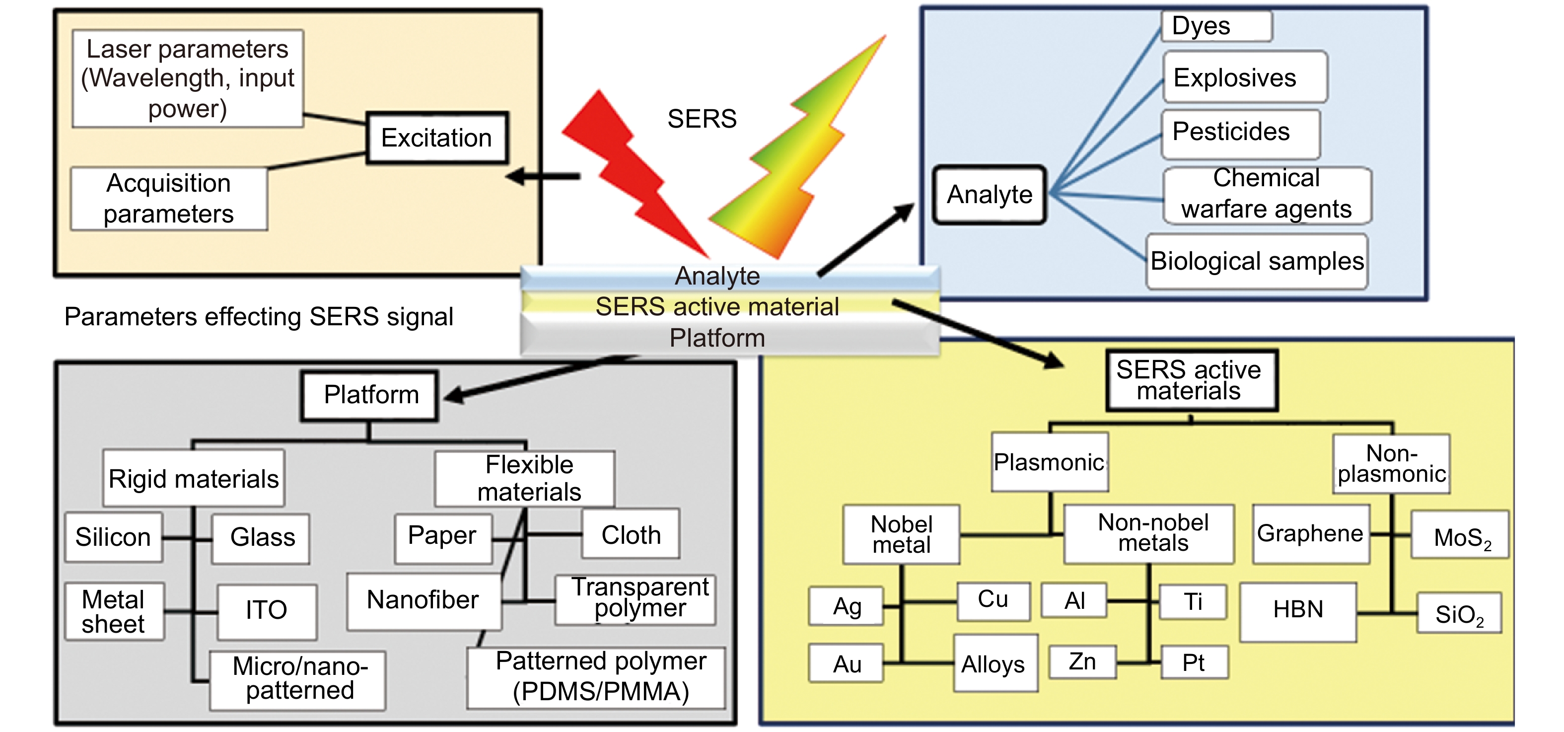
Figure 1.
A schematic depicting the various parameters influencing the SERS signal.
-
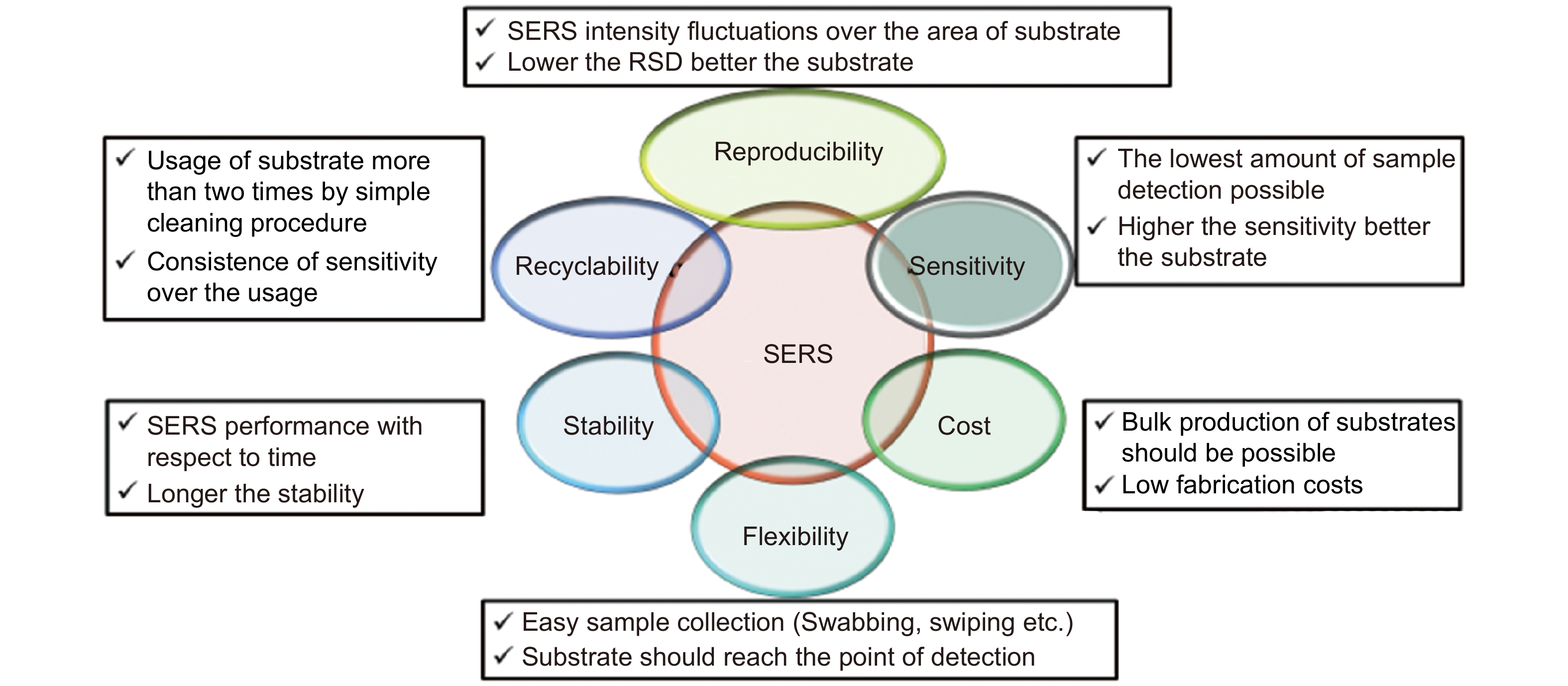
Figure 2.
The ideal requirements of SERS substrates are summarized in this schematic.
-
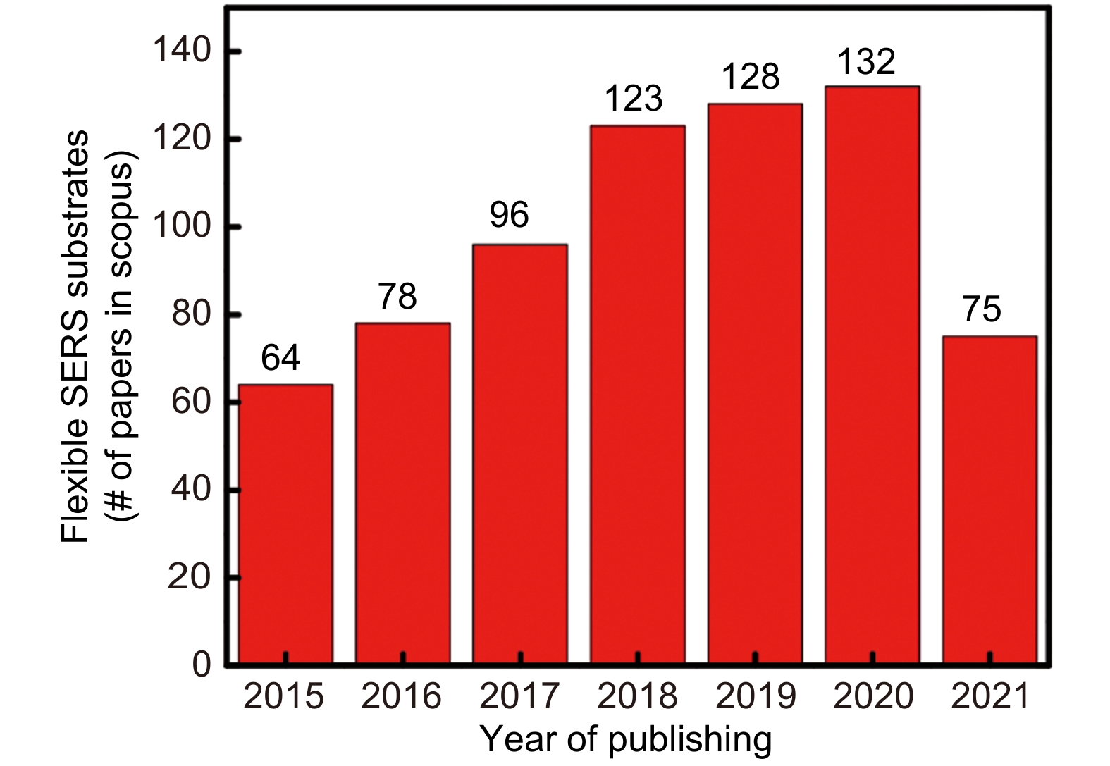
Figure 3.
Year wise publications on flexible SERS substrates obtained through a search in SCOPUS.
-
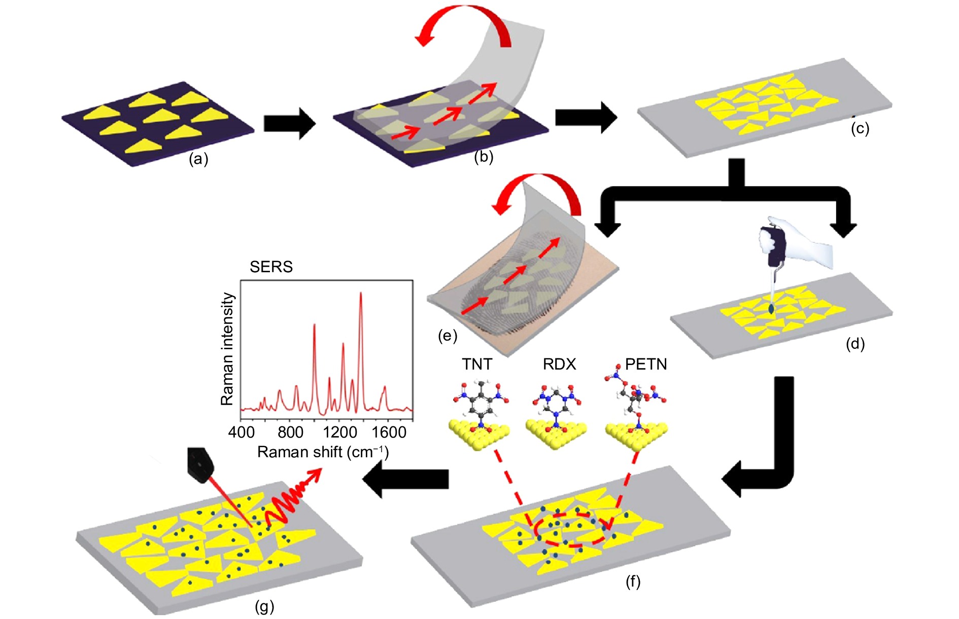
Figure 4.
Explosive trace detection using flexible SERS substrates detection of TNT, RDX, and PETN using self-assembly triangular nanoprisms on adhesive tape. Figure reproduced with permission from ref.58, Royal Society of Chemistry.
-
Figure 5.
Various fabrication techniques used for paper-based SERS substrates. (a) Vapor deposition. (b) Inkjet printing. (c) Dipping. (d) Pen-on-paper. (e) Drop-casting on hydrophilic wells. (f) Self assembling. (g) In-situ reduction. Figure reproduced with permission from: (a) ref.77, (b) ref.72, The Royal Society of Chemistry; ref.71, American Chemical Society; (d) ref.73, John Wiley and Sons; (e) ref.80, Springer Nature; (f) ref.84, (g) ref.79, American Chemical Society.
-
Figure 6.
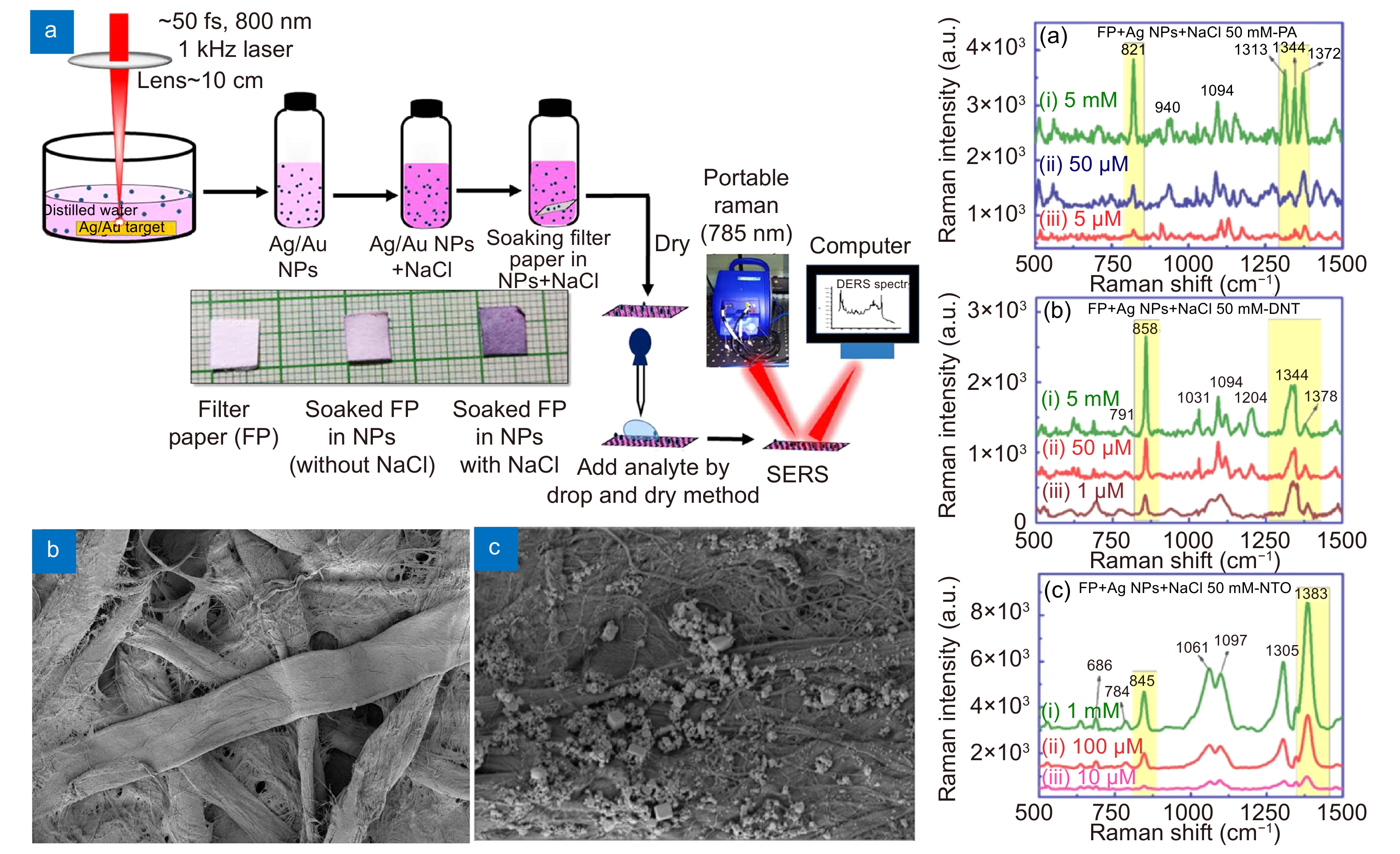
Filter paper based SERS substrate by aggregated Ag/Au NPs for explosive molecule detection (Left side) (a) schematic of substrate preparation (b) and (c) FESEM images of bare filter and aggregated Ag NPs (Right side) SERS spectra of (a) PA (b) DNT (c) NTO using FP with optimized aggregated Ag NPs. Figure reproduced with permission from ref.94, American Chemical Society.
-
Figure 7.
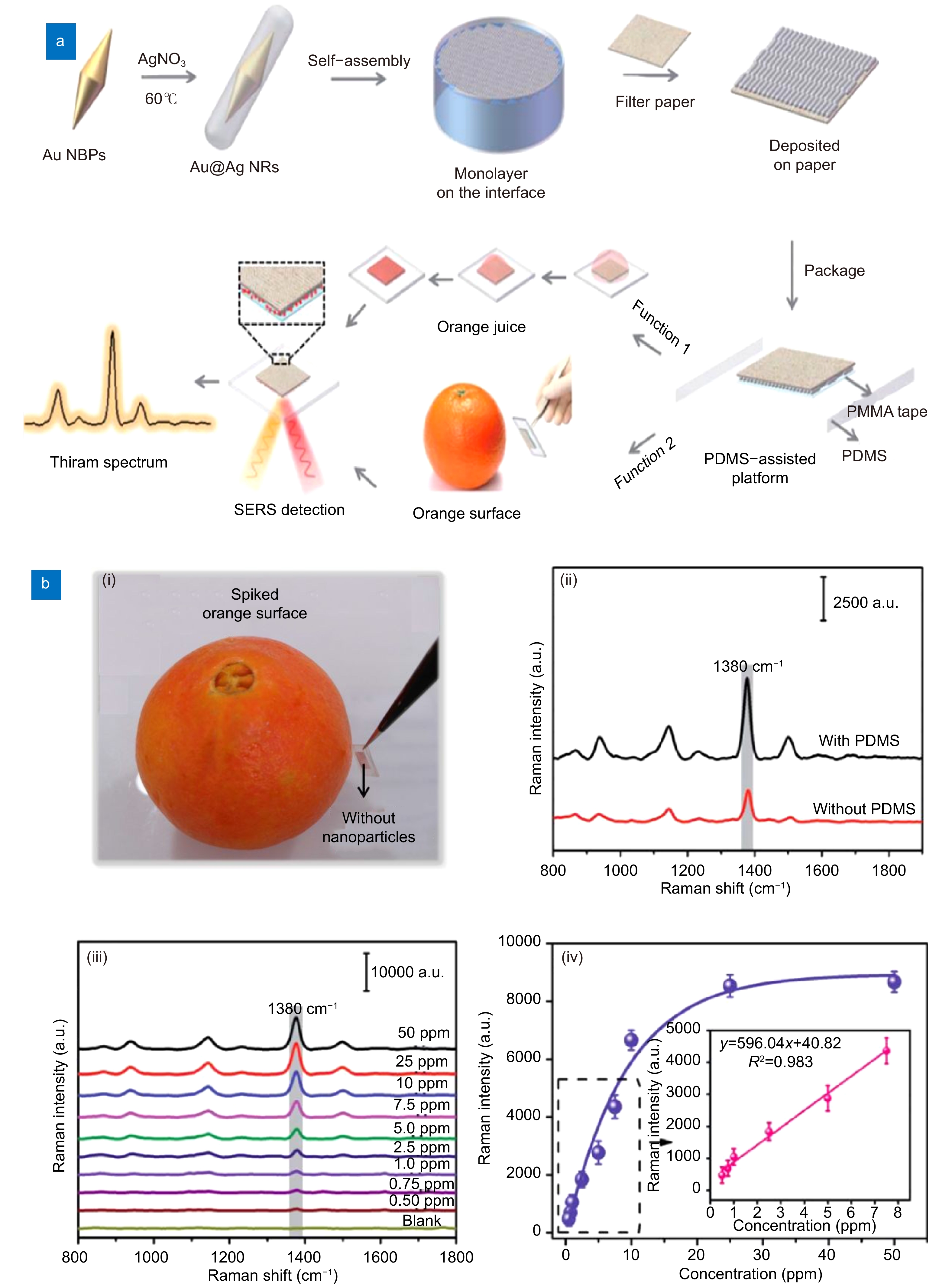
(a) A schematic of the synthesis of dual-functional PDMS-assisted paper-based SERS platform. (b) (i) The photograph of a sample collection from orange surface. (ii) A comparison of SERS spectra of CV with and without PDMS. (iii) SERS spectra of different concentrations of thiram (0.5−50 ppm). (iv) The peak intensity at 1380 cm−1 of thiram in orange juice as a function of the spiked sample concentration. Figure reproduced with permission from ref.95, Royal Society of Chemistry.
-
Figure 8.
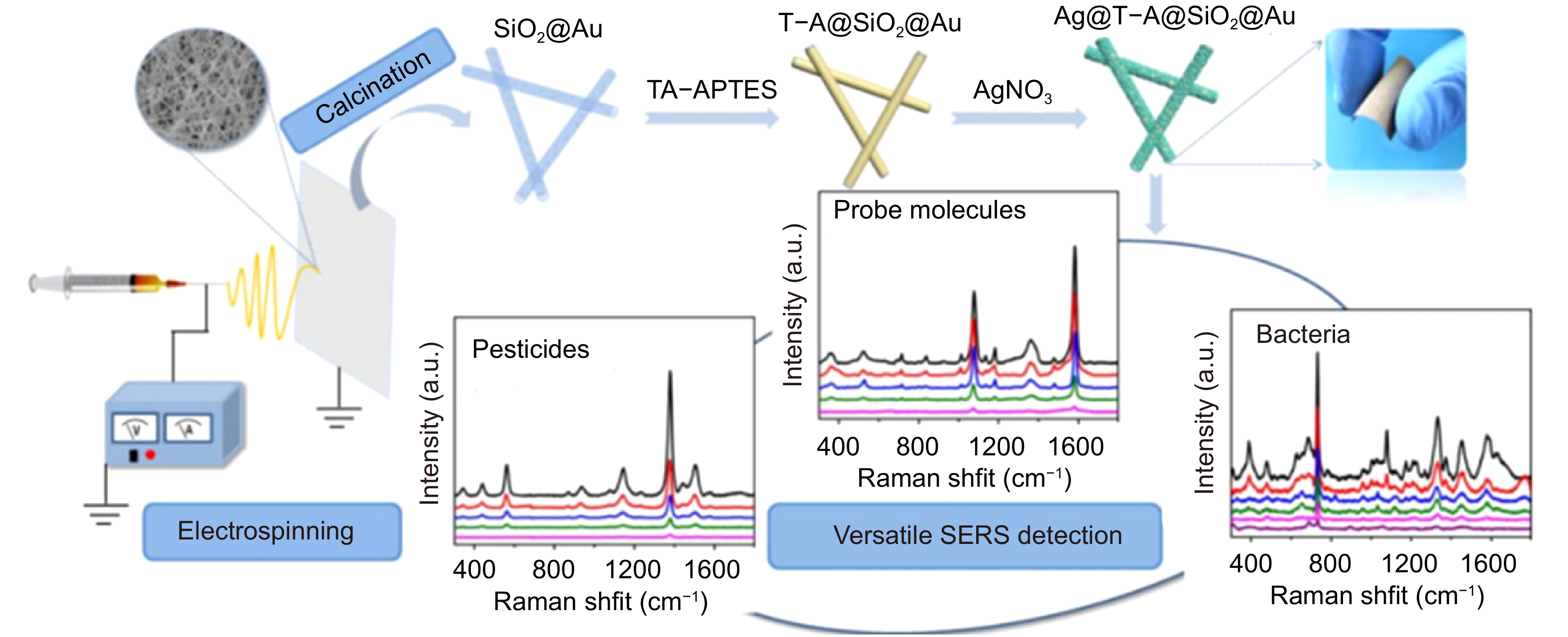
Fabrication of flexible SERS substrates for Ag@T-A@SiO2-Au nanofibrous substrates. Figure reproduced with permission from ref.100, under a Creative Commons Attribution 4.0 International License.
-
Figure 9.
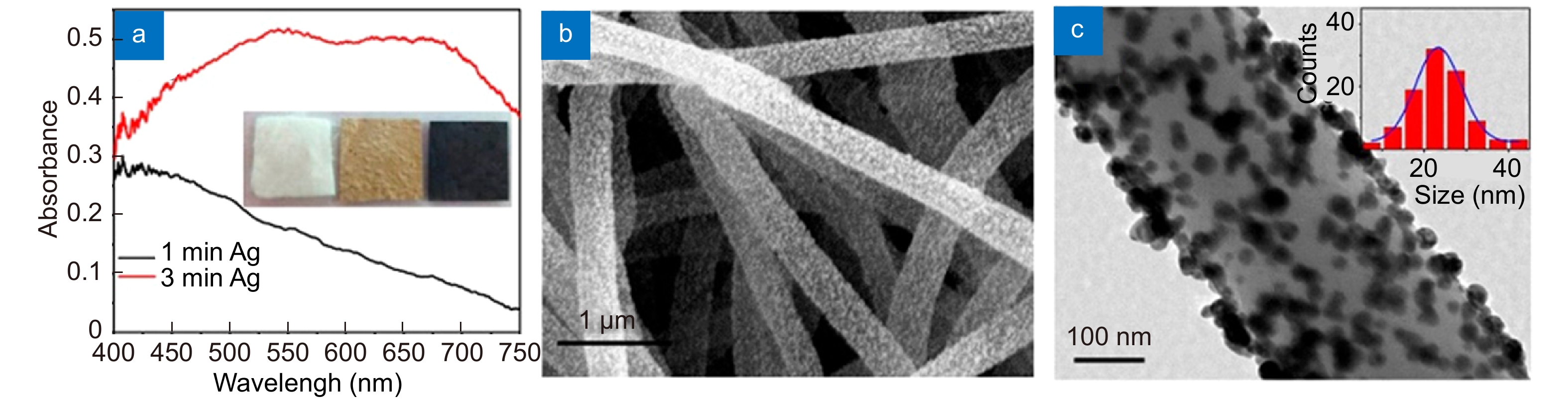
(a) Reflectance spectra of the ASFPAN nanofibrous membranes with Ag NPs; Photographs of three nanofibrous membranes (PAN, ASFPAN, and ASFPAN-Ag NPs) are shown in the inset. (b) SEM image and (c) TEM image of ASFPAN nanofibers (3 min). Inset in (c) shows the size distribution of Ag NPs.107, American Chemical Society.
-
Figure 10.
(a) Schematic diagram representing the fabrication process of Au covered polymer nanostructure arrays using roll-to-roll ultraviolet nanoimprint lithography (R2R UV-NIL) technique (b) and (c) SERS spectra of R6G from 30 nm Au coating flexible substrate at different bending angles and bending cycles, respectively. Figure reproduced with permission from ref.114, under a Creative Commons Attribution 4.0 International License.
-
Figure 11.
(a) Schematic of flexible non-woven fabric based substrate and the (b) SERS spectra of carbyl on apples, oranges, and bananas surfaces. Figure reproduced with permission from ref.129, under a Creative Commons Attribution 4.0 International License.


