
 E-mail Alert
 RSS
RSS2018 Vol. 45, No. 7
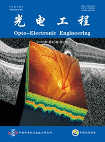
Cover Story:He J T, Chen M H, Jia W Y, et al. Segmentation of diabetic macular edema in OCT retinal images[J]. Opto-Electronic Engineering, 2018, 45(7): 170605;DOI: 10.12086/oee.2018.170605
Although ophthalmologists can diagnose patients with cystoid edema through OCT images, quantitative analysis of the lesion area is very difficult. The doctor needs to manually mark edema areas are marked on the OCT section of the fundus and are prone to errors. In addition, the OCT fundus data for each patient is very large. These works are undoubtedly time-consuming and labor-intensive. Research Team of Associate Professor Chen Minghui in Institute of Biomedical optics & Optometry, Shanghai Institute for Minimally Invasive Therapy, University of Shanghai for Science and Technology, long-term research in optical coherence tomography and application in biomedical optics. A method for segmenting diabetes in OCT images was proposed by the team. The method of macular edema can complete the segmentation with a relatively small amount of calculation and at a faster speed, and can segment a smaller edema region.

-
{{article.year}}, {{article.volume}}({{article.issue}}): {{article.fpage | processPage:article.lpage:6}}. doi: {{article.doi}}{{article.articleStateNameEn}}, Published online {{article.preferredDate | date:'dd MMMM yyyy'}}, doi: {{article.doi}}{{article.articleStateNameEn}}, Accepted Date {{article.acceptedDate | date:'dd MMMM yyyy'}}
Prof. Xiangang Luo


