
 E-mail Alert
 RSS
RSS
| Citation: |
Fei Qin, Xiangping Li, Minghui Hong. From super-osciallatory lens to super-critical lens: surpassing the diffraction limit via light field modulation[J]. Opto-Electronic Engineering, 2017, 44(8): 757-771. doi: 10.3969/j.issn.1003-501X.2017.08.001

|
From super-osciallatory lens to super-critical lens: surpassing the diffraction limit via light field modulation
-
Abstract
Super-oscillatory lens (SOL) and super-critical lens (SCL) are the typical representatives of planar metalens which could achieve sub-diffractive focusing and imaging in far field by means of light field modulation. Through precisely modulating the interference effect of each diffractive unit, the electromagnetic wave could be oscillated faster than its maximum frequency components in a certain region of the target plane, and then the focal spot size is controllable in lateral and longitudinal directions. Compared with the traditional optical lens, the planar metalens is much more attractive in the fields of diffractive optics and nanophotonics due to its distinct advantages of powerful focusing capabilities, compact configuration, higher design freedom and the integratable properties, etc. In this review, we briefly introduce the field modulation mechanism and design principle of planar metalens. The research advances of the super-oscillatory lens and super-critical lens, as well as their applications in far-field label-free super-resolution imaging, are discussed in detail. In addition, a perspective about the future outlook of planar metalens is summarized. Since the planar metalens has powerful capability in manipulating the light field, the rapid development in various applications would be gradually realized in the near future. -

-
References
[1] Abbe E. A contribution to the theory of the microscope and the nature of microscopic vision[C]//Proceedings of the Bristol Naturalists' Society, 1874, 1: 200-261. [2] Lord Rayleigh F R S. XII. On the manufacture and theory of diffraction-gratings[J]. Philosophical Magazine, 1874, 47(310): 81-93. [3] Airy G B. On the diffraction of an object-glass with circular aperture[J]. Transactions of the Cambridge Philosophical Society, 1835, 5: 283-291. [4] Hao Xiang, Kuang Cuifang, Gu Zhaotai, et al. From microscopy to nanoscopy via visible light[J]. Light Science & Applications, 2013, 2: e108. [5] Schmidt D A, Kopf I, Bründermann E. A matter of scale: from far-field microscopy to near-field nanoscopy[J]. Laser & Photonics Reviews, 2012, 6(3): 296-332. [6] Zeng Zhipeng, Xi Peng. Advances in three-dimensional super-resolution nanoscopy[J]. Microscopy Research and Technique, 2016, 79(10): 893-898. doi: 10.1002/jemt.v79.10 [7] Hell S W. Toward fluorescence nanoscopy[J]. Nature Biotechnology, 2003, 21(11): 1347-1355. doi: 10.1038/nbt895 [8] Hell S W. Far-field optical nanoscopy[J]. Science, 2007, 316(5828): 1153-1158. doi: 10.1126/science.1137395 [9] Wang H, Sheppard C J R, Ravi K, et al. Fighting against diffraction: apodization and near field diffraction structures[J]. Laser & Photonics Reviews, 2012, 6(3): 354-392. [10] Xie Xiangsheng, Chen Yongzhu, Yang Ken, et al. Harnessing the point-spread function for high-resolution far-field optical microscopy[J]. Physical Review Letters, 2014, 113(26): 263901. doi: 10.1103/PhysRevLett.113.263901 [11] Yang Xusan, Xie Hao, Alonas E, et al. Mirror-enhanced super-resolution microscopy[J]. Light: Science & Applications, 2016, 5: e16134. [12] Wang Wenhui, Gu Junnan, He Ting, et al. Optical super-resolution microscopy and its applications in nano-catalysis[J]. Nano Research, 2015, 8(2): 441-455. doi: 10.1007/s12274-015-0709-y [13] Synge E H. XXXVIII. A suggested method for extending microscopic resolution into the ultra-microscopic region[J]. The London, Edinburgh, and Dublin Philosophical Magazine and Journal of Science, 1928, 6(35): 356-362. doi: 10.1080/14786440808564615 [14] Betzig E, Lewis A, Harootunian A, et al. Near field scanning optical microscopy (NSOM)[J]. Biophysical Journal, 1986, 49(1): 269-279. doi: 10.1016/S0006-3495(86)83640-2 [15] Bek A, Vogelgesang R, Kern K. Apertureless scanning near field optical microscope with sub-10nm resolution[J]. Review of Scientific Instruments, 2006, 77(4): 043703. doi: 10.1063/1.2190211 [16] Pendry J B. Negative refraction makes a perfect lens[J]. Physical Review Letters, 2000, 85(18): 3966-3969. doi: 10.1103/PhysRevLett.85.3966 [17] Liu Zhaowei, Durant S, Lee H, et al. Far-field optical superlens[J]. Nano Letters, 2007, 7(2): 403-408. doi: 10.1021/nl062635n [18] Zhang Xiang, Liu Zhaowei. Superlenses to overcome the diffraction limit[J]. Nature Materials, 2008, 7(6): 435-441. doi: 10.1038/nmat2141 [19] Kawata S, Inouye Y, Verma P. Plasmonics for near-field nano-imaging and superlensing[J]. Nature Photonics, 2009, 3(7): 388-394. doi: 10.1038/nphoton.2009.111 [20] Fang N, Lee H, Sun Cheng, et al. Sub-diffraction-limited optical imaging with a silver superlens[J]. Science, 2005, 308(5721): 534-537. doi: 10.1126/science.1108759 [21] Taubner T, Korobkin D, Urzhumov Y, et al. Near-field micros-copy through a SiC superlens[J]. Science, 2006, 313(5793): 1595. doi: 10.1126/science.1131025 [22] Wang Zengbo, Guo Wei, Li Lin, et al. Optical virtual imaging at 50 nm lateral resolution with a white-light nanoscope[J]. Nature Communications, 2011, 2: 218. doi: 10.1038/ncomms1211 [23] Yan Yinzhou, Li Lin, Feng Chao, et al. Microsphere-coupled scanning laser confocal nanoscope for sub-diffraction-limited imaging at 25 nm lateral resolution in the visible spectrum[J]. ACS Nano, 2014, 8(2): 1809-1816. doi: 10.1021/nn406201q [24] Allen K W, Farahi N, Li Yangcheng, et al. Super-resolution microscopy by movable thin-films with embedded microspheres: Resolution analysis[J]. Annalen der Physik, 2015, 527(7-8): 513-522. doi: 10.1002/andp.v527.7-8 [25] Lee S, Li Lin, Wang Zengbo, et al. Immersed transparent microsphere magnifying sub-diffraction-limited objects[J]. Applied Optics, 2013, 52(30): 7265-7270. doi: 10.1364/AO.52.007265 [26] Darafsheh A, Walsh G F, Dal Negro L, et al. Optical super-resolution by high-index liquid-immersed microspheres[J]. Applied Physics Letters, 2012, 101(14): 141128. doi: 10.1063/1.4757600 [27] Li Lin, Guo Wei, Yan Yinzhou, et al. Label-free super-resolution imaging of adenoviruses by submerged microsphere optical nanoscopy[J]. Light: Science & Applications, 2013, 2: e104. [28] Yang Hui, Trouillon R, Huszka G, et al. Super-resolution imaging of a dielectric microsphere is governed by the waist of its photonic nanojet[J]. Nano Letters, 2016, 16(8): 4862-4870. doi: 10.1021/acs.nanolett.6b01255 [29] Allen K W, Farahi N, Li Yangcheng, et al. Overcoming the diffraction limit of imaging nanoplasmonic arrays by micro-spheres and microfibers[J]. Optics Express, 2015, 23(19): 24484-24496. doi: 10.1364/OE.23.024484 [30] Wu M X, Huang B J, Chen R, et al. Modulation of photonic nanojets generated by microspheres decorated with concentric rings[J]. Optics Express, 2015, 23(15): 20096-20103. doi: 10.1364/OE.23.020096 [31] Wu Mengxue, Chen Rui, Ling Jinzhong, et al. Creation of a longitudinally polarized photonic nanojet via an engineered microsphere[J]. Optics Letters, 2017, 42(7): 1444-1447. doi: 10.1364/OL.42.001444 [32] Fan Wen, Yan Bing, Wang Zengbo, et al. Three-dimensional all-dielectric metamaterial solid immersion lens for subwavelength imaging at visible frequencies[J]. Science Advances, 2016, 2(8): e1600901. [33] Li Jinxing, Liu Wenjuan, Li Tianlong, et al. Swimming microrobot optical nanoscopy[J]. Nano Letters, 2016, 16(10): 6604-6609. doi: 10.1021/acs.nanolett.6b03303 [34] Luk'yanchuk B S, Paniagua-Domínguez R, Minin I, et al. Refractive index less than two: photonic nanojets yesterday, today and tomorrow[J]. Optical Materials Express, 2017, 7(6): 1820-1847. doi: 10.1364/OME.7.001820 [35] Liu Hong, Wang Bing, Ke Lin, et al. High aspect subdiffraction-limit photolithography via a silver superlens[J]. Nano Letters, 2012, 12(3): 1549-1554. doi: 10.1021/nl2044088 [36] Liu Hong, Wang Bing, Ke Lin, et al. High contrast superlens lithography engineered by loss reduction[J]. Advanced Functional Materials, 2012, 22(18): 3777-3783. doi: 10.1002/adfm.v22.18 [37] Srituravanich W, Fang N, Sun Cheng, et al. Plasmonic nanolithography[J]. Nano Letters, 2004, 4(6): 1085-1088. doi: 10.1021/nl049573q [38] Liu Zhaowei, Wei Qihuo, Zhang Xiang. Surface plasmon interference nanolithography[J]. Nano Letters, 2005, 5(5): 957-961. doi: 10.1021/nl0506094 [39] Luo Xiangang, Ishihara T. Surface plasmon resonant interference nanolithography technique[J]. Applied Physics Letters, 2004, 84(23): 4780. doi: 10.1063/1.1760221 [40] Gao Ping, Yao Na, Wang Changtao, et al. Enhancing aspect profile of half-pitch 32 nm and 22 nm lithography with plasmonic cavity lens[J]. Applied Physics Letters, 2015, 106(9): 093110. doi: 10.1063/1.4914000 [41] Gustafsson M G L. Surpassing the lateral resolution limit by a factor of two using structured illumination microscopy[J]. Journal of Microscopy, 2000, 198(2): 82-87. doi: 10.1046/j.1365-2818.2000.00710.x [42] Gustafsson M G L. Nonlinear structured-illumination microscopy: Wide-field fluorescence imaging with theoretically unlimited resolution[J]. Proceedings of the National Academy of Sciences of the United States of America, 2005, 102(37): 13081-13086. doi: 10.1073/pnas.0406877102 [43] Allen J R, Ross S T, Davidson M W. Structured illumination microscopy for superresolution[J]. Chemphyschem, 2014, 15(4): 566-576. doi: 10.1002/cphc.201301086 [44] Rust M J, Bates M, Zhuang Xiaowei. Sub-diffraction-limit imaging by stochastic optical reconstruction microscopy (STORM)[J]. Nature Methods, 2006, 3(10): 793-796. doi: 10.1038/nmeth929 [45] Bates M, Huang Bo, Dempsey G T, et al. Multicolor super-resolution imaging with photo-switchable fluorescent probes[J]. Science, 2007, 317(5845): 1749-1753. doi: 10.1126/science.1146598 [46] Huang Bo, Wang Wenqin, Bates M, et al. Three-dimensional super-resolution imaging by stochastic optical reconstruction microscopy[J]. Science, 2008, 319(5864): 810-813. doi: 10.1126/science.1153529 [47] Dempsey G T, Bates M, Kowtoniuk W E, et al. Photoswitching mechanism of cyanine dyes[J]. Journal of the American Chemical Society, 2009, 131(151): 18192-18193. [48] Betzig E, Patterson G H, Sougrat R, et al. Imaging intracellular fluorescent proteins at nanometer resolution[J]. Science, 2006, 313(5793): 1642-1645. doi: 10.1126/science.1127344 [49] Shroff H, Galbraith C G, Galbraith J A, et al. Live-cell photoactivated localization microscopy of nanoscale adhesion dynamics[J]. Nature Methods, 2008, 5(5): 417-423. doi: 10.1038/nmeth.1202 [50] Planchon T A, Gao Liang, Milkie D E, et al. Rapid three-dimensional isotropic imaging of living cells using Bessel beam plane illumination[J]. Nature Methods, 2011, 8(5): 417-423. doi: 10.1038/nmeth.1586 [51] Hell S W, Wichmann J. Breaking the diffraction resolution limit by stimulated emission: stimulated-emission-depletion fluorescence microscopy[J]. Optics Letters, 1994, 19(11): 780-782. doi: 10.1364/OL.19.000780 [52] Willig K I, Rizzoli S O, Westphal V, et al. STED microscopy reveals that synaptotagmin remains clustered after synaptic vesicle exocytosis[J]. Nature, 2006, 440(7086): 935-939. doi: 10.1038/nature04592 [53] Bretschneider S, Eggeling C, Hell S W. Breaking the diffraction barrier in fluorescence microscopy by optical shelving[J]. Physical Review Letters, 2007, 98(21): 218103. doi: 10.1103/PhysRevLett.98.218103 [54] Willig K I, Harke B, Medda R, et al. STED microscopy with continuous wave beams[J]. Nature Methods, 2007, 4(11): 915-918. doi: 10.1038/nmeth1108 [55] Rittweger E, Han K Y, Irvine S E, et al. STED microscopy reveals crystal colour centres with nanometric resolution[J]. Nature Photonics, 2009, 3(3): 144-147. doi: 10.1038/nphoton.2009.2 [56] Grotjohann T, Testa I, Leutenegger M, et al. Diffraction-unlimited all-optical imaging and writing with a photochromic GFP[J]. Nature, 2011, 478(7368): 204-208. doi: 10.1038/nature10497 [57] Berning S, Willig K I, Steffens H, et al. Nanoscopy in a living mouse brain[J]. Science, 2012, 335(6068): 551. doi: 10.1126/science.1215369 [58] Hanne J, Falk H J, Görlitz F, et al. STED nanoscopy with fluorescent quantum dots[J]. Nature Communications, 2015, 6: 7127. doi: 10.1038/ncomms8127 [59] Hell S W, Sahl S J, Bates M, et al. The 2015 super-resolution microscopy roadmap[J]. Journal of Physics D: Applied Physics, 2015, 48(44): 443001. doi: 10.1088/0022-3727/48/44/443001 [60] Di Francia G T. Super-gain antennas and optical resolving power[J]. Nuovo Cimento, 1952, 9(S3): 426-438. doi: 10.1007/BF02903413 [61] Liu Tao, Tan Jiubin, Liu Jian, et al. Creation of subwavelength light needle, equidistant multi-focus, and uniform light tunnel[J]. Journal of Modern Optics, 2013, 60(5): 378-381. doi: 10.1080/09500340.2013.778343 [62] Liu Tao, Shen Tong, Yang Shuming, et al. Subwavelength focusing by binary multi-annular plates: design theory and experiment[J]. Journal of Optics, 2015, 17(3): 035610. doi: 10.1088/2040-8978/17/3/035610 [63] Liu Tao, Liu Jian, Zhang He, et al. Efficient optimization of super-oscillatory lens and transfer function analysis in confocal scanning microscopy[J]. Optics Communications, 2014, 319: 31-35. doi: 10.1016/j.optcom.2013.12.054 [64] Sheppard C J R, Choudhury A. Annular pupils, radial polarization, and superresolution[J]. Applied Optics, 2004, 43(22): 4322-4327. doi: 10.1364/AO.43.004322 [65] Davis B J, Karl W C, Swan A K, et al. Capabilities and limitations of pupil-plane filters for superresolution and image enhancement[J]. Optics Express, 2004, 12(17): 4150-4156. doi: 10.1364/OPEX.12.004150 [66] Huang Kun, Li Yongping. Realization of a subwavelength focused spot without a longitudinal field component in a solid immersion lens-based system[J]. Optics Letters, 2011, 36(18): 3536-3538. doi: 10.1364/OL.36.003536 [67] Huang Kun, Shi Peng, Kang Xueliang, et al. Design of DOE for generating a needle of a strong longitudinally polarized field[J]. Optics Letters, 2010, 35(7): 965-967. doi: 10.1364/OL.35.000965 [68] Wang Haifeng, Shi Luping, Lukyanchuk B, et al. Creation of a needle of longitudinally polarized light in vacuum using binary optics[J]. Nature Photonics, 2008, 2(8): 501-505. doi: 10.1038/nphoton.2008.127 [69] Berry M V. Exact nonparaxial transmission of subwavelength detail using superoscillations[J]. Journal of Physics A: Mathematical and Theoretical, 2013, 46(20): 205203. doi: 10.1088/1751-8113/46/20/205203 [70] Berry M V, Popescu S. Evolution of quantum superoscillations and optical superresolution without evanescent waves[J]. Journal of Physics A: Mathematical and Theoretical, 2006, 39(22): 6965-6977. doi: 10.1088/0305-4470/39/22/011 [71] Huang Fumin, Zheludev N, Chen Yifang, et al. Focusing of light by a nanohole array[J]. Applied Physics Letters, 2007, 90(9): 091119. doi: 10.1063/1.2710775 [72] Roy T, Rogers E T F, Yuan Guanghui, et al. Point spread function of the optical needle super-oscillatory lens[J]. Applied Physics Letters, 2014, 104(23): 231109. doi: 10.1063/1.4882246 [73] Huang Fumin, Chen Yifang, de Abajo F J G, et al. Optical super-resolution through super-oscillations[J]. Journal of Optics A: Pure and Applied Optics, 2007, 9(9): S285-S288. doi: 10.1088/1464-4258/9/9/S01 [74] Rogers E T F, Zheludev N I. Optical super-oscillations: sub-wavelength light focusing and super-resolution imaging[J]. Journal of Optics, 2013, 15(9): 094008. doi: 10.1088/2040-8978/15/9/094008 [75] Rogers E T F, Savo S, Lindberg J, et al. Super-oscillatory optical needle[J]. Applied Physics Letters, 2013, 102(3): 031108. doi: 10.1063/1.4774385 [76] Yuan Guanghui, Rogers E T F, Zheludev N I. Achromatic super-oscillatory lenses with sub-wavelength focusing[J]. Light: Science & Applications, 2017, 6: e17036. [77] Yuan Guanghui, Vezzoli S, Altuzarra C, et al. Quantum super-oscillation of a single photon[J]. Light: Science & Applications, 2016, 5: e16127. [78] Huang Fumin, Kao T S, Fedotov V A, et al. Nanohole array as a lens[J]. Nano Letters, 2008, 8(8): 2469-2472. doi: 10.1021/nl801476v [79] Huang Fumin, Zheludev N I. Super-resolution without evanescent waves[J]. Nano Letters, 2009, 9(3): 1249-1254. doi: 10.1021/nl9002014 [80] Rogers E T F, Lindberg J, Roy T, et al. A super-oscillatory lens optical microscope for subwavelength imaging[J]. Nature Materials, 2012, 11(5): 432-435. doi: 10.1038/nmat3280 [81] Wang Qian, Rogers E T F, Gholipour B, et al. Optically reconfigurable metasurfaces and photonic devices based on phase change materials[J]. Nature Photonics, 2015, 10(1): 60-65. [82] Zheludev N I. What diffraction limit?[J]. Nature Materials, 2008, 7(6): 420-422. doi: 10.1038/nmat2163 [83] Roy T, Rogers E T F, Zheludev N I. Sub-wavelength focusing meta-lens[J]. Optics Express, 2013, 21(6): 7577-7582. doi: 10.1364/OE.21.007577 [84] Yuan Guanghui, Rogers E T F, Roy T, et al. Planar su-per-oscillatory lens for sub-diffraction optical needles at violet wavelengths[J]. Scientific Reports, 2014, 4: 6333. [85] Huang Kun, Ye Huapeng, Teng Jinghua, et al. Optimiza-tion-free superoscillatory lens using phase and amplitude masks[J]. Laser & Photonics Reviews, 2014, 8(1): 152-157. [86] Ye Huapeng, Qiu Chengwei, Huang Kun, et al. Creation of a longitudinally polarized subwavelength hotspot with an ultra-thin planar lens: vectorial Rayleigh-Sommerfeld method[J]. Laser Physics Letters, 2013, 10(6): 065004. doi: 10.1088/1612-2011/10/6/065004 [87] Qin Fei, Huang Kun, Wu Jianfeng, et al. A supercritical lens optical label-free microscopy: sub-diffraction resolution and ultra-long working distance[J]. Advanced Materials, 2017, 29(8): 1602721. doi: 10.1002/adma.201602721 [88] Wang Jun, Qin Fei, Zhang Daohua, et al. Subwavelength superfocusing with a dipole-wave-reciprocal binary zone plate[J]. Applied Physics Letters, 2013, 102(6): 061103. doi: 10.1063/1.4791581 [89] Tang Dongliang, Wang Changtao, Zhao Zeyu, et al. Ultrabroadband superoscillatory lens composed by plasmonic metasurfaces for subdiffraction light focusing[J]. Laser & Photonics Reviews, 2015, 9(6): 713-719. [90] Qin Fei, Hong Minghui. Breaking the diffraction limit in far field by planar Metalens[J]. Science China Physics, Mechanics & Astronomy, 2017, 60(4): 044231. [91] Qin Fei, Huang Kun, Wu Jianfeng, et al. Shaping a subwavelength needle with ultra-long focal length by focusing azimuthally polarized light[J]. Scientific Reports, 2015, 5: 9977. doi: 10.1038/srep09977 [92] Wang Changtao, Tang Dongliang, Wang Yanqin, et al. Super-resolution optical telescopes with local light diffraction shrinkage[J]. Scientific Reports, 2015, 5: 18485. [93] Richards B, Wolf E. Electromagnetic diffraction in optical systems II. structure of the image field in an aplanatic system[J]. Proceedings of the Royal Society A: Mathematical, Physical and Engineering Sciences, 1959, 253(1274): 358-379. doi: 10.1098/rspa.1959.0200 [94] Lerman G M, Yanai A, Levy U. Demonstration of nanofocusing by the use of plasmonic lens illuminated with radially polarized light[J]. Nano Letters, 2009, 9(5): 2139-2143. doi: 10.1021/nl900694r [95] Wilson T, Massoumian F, Juškaitis R. Generation and focusing of radially polarized electric fields[J]. Optical Engineering, 2003, 42(11): 3088-3089. doi: 10.1117/1.1618816 [96] Huang Kun, Shi Peng, Cao G W, et al. Vector-vortex Bes-sel-Gauss beams and their tightly focusing properties[J]. Optics Letters, 2011, 36(6): 888-890. doi: 10.1364/OL.36.000888 [97] Li Xiangping, Cao Yaoyu, Gu Min. Superresolution-focal-vol-ume induced 3.0 Tbytes/disk capacity by focusing a radially polarized beam[J]. Optics Letters, 2011, 36(13): 2510-2512. doi: 10.1364/OL.36.002510 [98] Li Xiangping, Venugopalan P, Ren Haoran, et al. Super-resolved pure-transverse focal fields with an enhanced energy density through focus of an azimuthally polarized first-order vortex beam[J]. Optics Letters, 2014, 39(20): 5961-5964. doi: 10.1364/OL.39.005961 [99] Zhan Qiwen. Cylindrical vector beams: from mathematical concepts to applications[J]. Advances in Optics and Photonics, 2009, 1(1): 1-57. doi: 10.1364/AOP.1.000001 [100] Youngworth K S, Brown T G. Focusing of high numerical aperture cylindrical-vector beams[J]. Optics Express, 2000, 7(2): 77-87. doi: 10.1364/OE.7.000077 [101] Dorn R, Quabis S, Leuchs G. Sharper focus for a radially polarized light beam[J]. Physical Review Letters, 2003, 91(23): 233901. doi: 10.1103/PhysRevLett.91.233901 [102] Liu Hong, Mehmood M Q, Huang Kun, et al. Twisted focusing of optical vortices with broadband flat spiral zone plates[J]. Advanced Optical Materials, 2014, 2(12): 1193-1198. doi: 10.1002/adom.201400315 [103] Huang Kun, Liu Hong, Garcia-Vidal F J, et al. Ultra-high-capacity non-periodic photon sieves operating in visible light[J]. Nature Communications, 2015, 6: 7059. doi: 10.1038/ncomms8059 [104] Wang Sicong, Li Xiangping, Zhou Jianying, et al. Ultralong pure longitudinal magnetization needle induced by annular vortex binary optics[J]. Optics Letters, 2014, 39(17): 5022-5025. doi: 10.1364/OL.39.005022 [105] Chen Gang, Wu Zhixiang, Yu Anping, et al. Generation of a sub-diffraction hollow ring by shaping an azimuthally polarized wave[J]. Scientific Reports, 2016, 6: 37776. doi: 10.1038/srep37776 [106] Yu Anping, Chen Gang, Zhang Zhihai, et al. Creation of sub-diffraction longitudinally polarized spot by focusing radially polarized light with binary phase lens[J]. Scientific Reports, 2016, 6: 38859. doi: 10.1038/srep38859 [107] Qin Fei, Ding Lu, Zhang Lei, et al. Hybrid bilayer plasmonic metasurface efficiently manipulates visible light[J]. Science Advances, 2016, 2(1): e1501168. doi: 10.1126/sciadv.1501168 [108] Zhang Lei, Mei Shengtao, Huang Kun, et al. Advances in full control of electromagnetic waves with metasurfaces[J]. Ad-vanced Optical Materials, 2016, 4(6): 818-833. doi: 10.1002/adom.v4.6 [109] Aieta F, Genevet P, Kats M A, et al. Aberration-free ultrathin flat lenses and axicons at telecom wavelengths based on plasmonic metasurfaces[J]. Nano Letters, 2012, 12(9): 4932-4936. doi: 10.1021/nl302516v [110] Devlin R C, Khorasaninejad M, Chen Weiting, et al. Broadband high-efficiency dielectric metasurfaces for the visible spectrum[J]. Proceedings of the National Academy of Sciences of the United States of America, 2016, 113(38): 10473-10478. doi: 10.1073/pnas.1611740113 [111] Aieta F, Kats M A, Genevet P, et al. Multiwavelength achromatic metasurfaces by dispersive phase compensation[J]. Science, 2015, 347(6228): 1342-1345. doi: 10.1126/science.aaa2494 [112] Khorasaninejad M, Chen Weiting, Devlin R C, et al. Metalenses at visible wavelengths: diffraction-limited focusing and subwavelength resolution imaging[J]. Science, 2016, 352(6290): 1190-1194. doi: 10.1126/science.aaf6644 [113] Yu Nanfang, Genevet P, Kats M A, et al. Light propagation with phase discontinuities: generalized laws of reflection and refraction[J]. Science, 2011, 334(6054): 333-337. doi: 10.1126/science.1210713 [114] Shen Yue, Luo Xiangang. Efficient bending and focusing of light beam with all-dielectric subwavelength structures[J]. Optics Communications, 2016, 366: 174-178. doi: 10.1016/j.optcom.2015.12.043 [115] Luo Xiangang. Principles of electromagnetic waves in metasurfaces[J]. Science China Physics, Mechanics & As-tronomy, 2015, 58(9): 594201. [116] Pu Mingbo, Li Xiong, Ma Xiaoliang, et al. Catenary optics for achromatic generation of perfect optical angular momentum[J]. Science Advances, 2015, 1(9): e1500396. doi: 10.1126/sciadv.1500396 [117] Zhao Xiaonan, Hu Jingpei, Lin Yu, et al. Ultra-broadband achromatic imaging with diffractive photon sieves[J]. Scientific Reports, 2016, 6: 28319. doi: 10.1038/srep28319 -
Overview

Improving the imaging resolution has always been one of the most important topics since the invention of optical microscope. Due to the fundamental laws of wave optics, the focusing and imaging resolution of traditional refraction and diffraction lenses are subject to the Rayleigh Criterion (0.61λ/NA), and the spatial resolution of optical microscopy is restricted to ~200 nm at visible light. Tremendous efforts have been made to fight against the diffraction limit in the past decades, and several novel approaches have been invented which could be categorized as near-field and far-field modes. For the near-field techniques, such as NSOM, superlens, hyperlens, microsphere lens, they always suffer from the challenges of near-field operation and small field of view, which make them not meet some requirements of practical applications. Although very high imaging resolution in far-field could be achieved by the fluorescence-based approaches, all these techniques have a common feature that is quite limited to biological domain because of the requirement to put dyes and fluorescence into objects. Therefore, the label-free technique for super-resolution imaging in far field is very important for general applications. Recent advance in this field is the development of planar metalens which could achieve sub-diffractive focusing and imaging in far field by means of light field modulation. Super-oscillatory lens (SOL) and super-critical lens (SCL) are the typical representatives of planar metalens. Through precisely modulating the interference effect of each diffractive unit, the focal spot size in a certain region of the target plane is controllable in lateral and longitudinal directions. Combined with the confocal technique, the label-free superresolution imaging could be realized in far field with purely non-invasive manners. Compared with the traditional optical lens, the planar metalens is much more attractive due to its distinct advantages of powerful focusing capabilities, compact configuration, higher design freedom and the integratable properties, etc. In this review, we briefly introduce the field modulation mechanism and design principle of the planar metalens. The research progress of the super-oscillatory lens and super-critical lens, as well as their applications in far-field label-free super-resolution imaging, is presented in detail. The advantages and limitations of that planar lens are compared and briefly discussed. A perspective about the future outlook of planar metalens is summarized. Since the planar metalens has a powerful capability in manipulating the light field, the rapid development in various applications would be gradually realized in the near future.
-
Access History

Export File
Citation
Fei Qin, Xiangping Li, Minghui Hong. From super-osciallatory lens to super-critical lens: surpassing the diffraction limit via light field modulation[J]. Opto-Electronic Engineering, 2017, 44(8): 757-771. doi: 10.3969/j.issn.1003-501X.2017.08.001
Format
Content
 DownLoad:

-
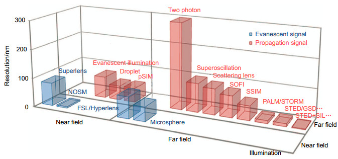
Figure 1.
Comparison between typical optical super-resolution techniques and their corresponding imaging resolutions[4].
-
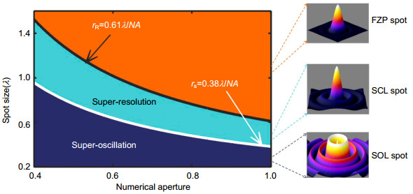
Figure 2.
The focal spot size of planar diffractive lens could be divided into three parts by Rayleigh (black) and super-oscillation (white) criterions, including sub-resolved (orange), super-resolution (cyan) and super-oscillation (dark blue). The insets in the right side are the field distributions of the focal spots for three typical diffractive lenses[85].
-
Figure 3.
The diffraction effect at a certain plane(z=20λ) for a single belt with different widths and numerical apertures. (a) Illustration of the diffraction effect of a single belt with its radius r0 and width △r. (b) Root-mean-square error (RMSE) between the diffracting intensity at the target plane and its corresponding zero-order Bessel function for different widths and radius of a single belt. (c), (d) The line profiles of the diffraction intensities at the positions A and B in Fig. 3(b), with its corresponding zero-order Bessel function with the same numerical apertures. (e) The dependence of the amplitude modulation coefficient on the width and radius of the single belt[85].
-
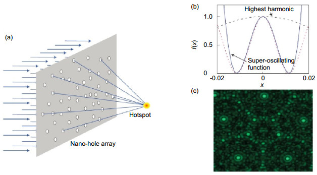
Figure 4.
(a) Generation of a sub-diffractive hotspot by nanoholes array in an opaque screen. (b) The comparison between the super-oscillating functions with its highest harmonic fourier component. (c) The experimental results of the subwavelength super-oscillating focal spot by a quasi-periodical holes array[82].
-
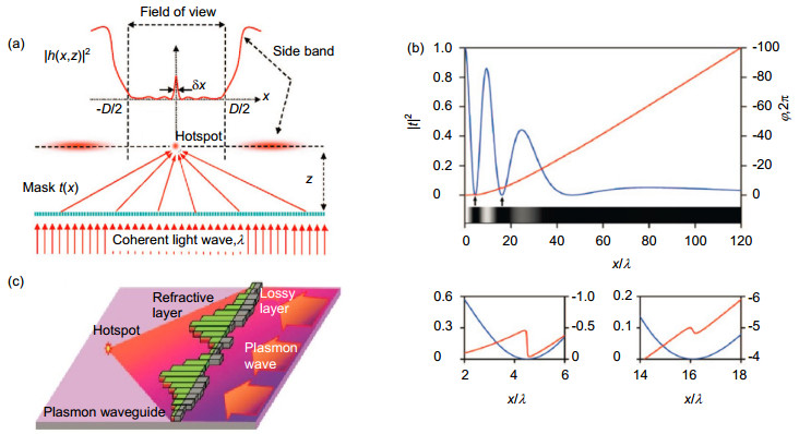
Figure 5.
(a) Schematic of the optical super-oscillation effect. (b) The intensity and phase profile of a transmission mask which could generate a subwavelength hotspot. (c) A possible configuration of a plasmonic focusing device for creating super-oscillation hotspot[79].
-
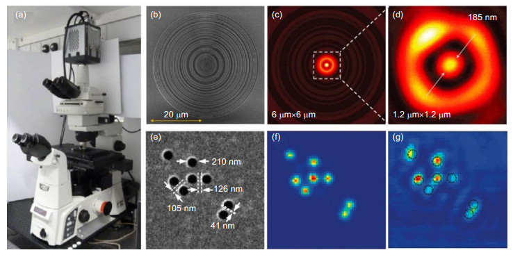
Figure 6.
(a) Photograph of the super-oscillatory microscope. (b) SEM image of the fabricated SOL. (c) The simulated energy distribution for the 640 nm wavelength SOL at the distance of 10.3 μm away from the lens plane. (d) Experimental focal spot with a FWHM of 185 nm. (e) SEM image of a hole array sample. (f) Simulated imaging result of the hole array sample by the SOL microscopy. (g) Experimental imaging result by the SOL microscopy[74, 80].
-
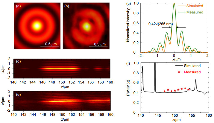
Figure 7.
Focusing effect of the 633 nm super-critical lens induced by the azimuthally polarized beam with vorticle phase. (a) Simulated energy distribution at the focal plane of z=150 μm. (b) Experimental recorded focal spot pattern. (c) Line profile of the intensity distribution for the simulated and measured focal spot. (d), (e) Simulated and experimental recorded optical needle formed in the range from z=140 μm~160 μm. (f) FWHM of the optical needle along the propagation direction[91].
-
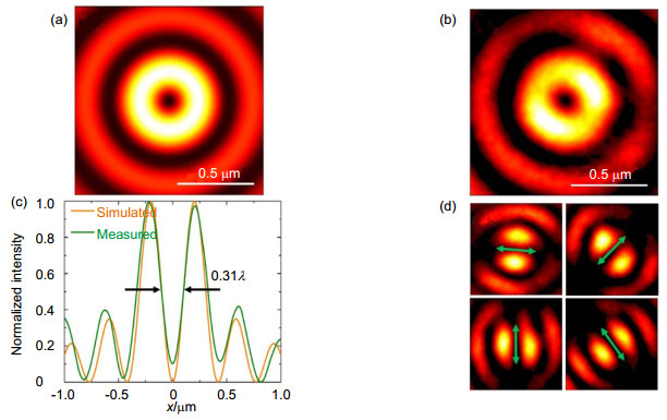
Figure 8.
(a), (b) The simulated and measured optical hollows created by the 633 nm SCL induced by azimuthally polarized beam. (c) Line profile of the intensity across the focal spot for the simulated and measured results. (d) Characterization of the polarized property of the optical hollow[91].
-
Figure 9.
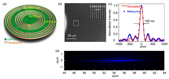
(a) Schematic configuration of the 405 nm supercritical lens. (b) SEM image of the fabricated 405 nm SCL. Inset is the zoom-in view of the dashed box region. (c) Line profile of the sub-diffractive focal spot under illumination of 405 nm circular polarized beam. (d) Experimental recorded intensity distribution of the sub-wavelength optical needle[87].
-
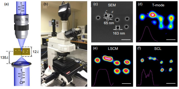
Figure 10.
(a) Schematic of the SCL microscopy. (b) The photograph of the SCL microscope system. (c) SEM of the nanoscale big dipper as the imaging specimen. (d) Imaging result by the normal transmission-mode microscopy. (e) Imaging results by the laser scanning confocal microscopy. (f) Imaging result by the 405 nm SCL microscopy[87].
-
Figure 11.
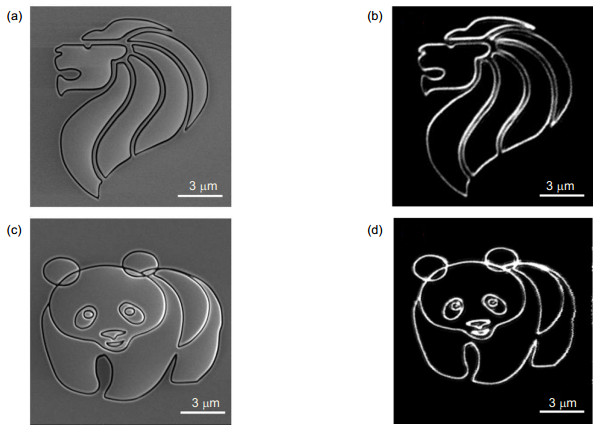
Large-scale non-periodic patterns imaged by the supercritical lens microscopy. (a), (c) The SEM images of fabricated samples with a size of 13.5 μm × 13.5 μm. (b), (d) The imaging results by the supercritical lens microscope[87].
-
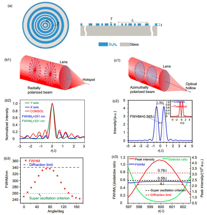
Figure 12.
(a) Schematic of the binary phase planar metalens. (b1)~(b3) Sub-diffractive focusing by the binary phase planar metalens under illumination of radial polarized beam. (c1)~(c3) Shaping subwavelength optical hollow with the binary phase planar metalens induced by azimuthally polarized beam[105, 106].
-
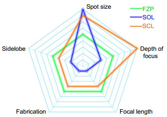
Figure 13.
Quality indices of three types of planar metalens. FZP, SOL and SCL refer to the fresnel zone plate, superoscillatory lens and supercritical lens, respectively.


