
 E-mail Alert
 RSS
RSS
| Citation: |
Zhang Xueying, Wang Jinsong, Huang Guolin, et al. Design of optical system for high accuracy imaging keratometry[J]. Opto-Electronic Engineering, 2019, 46(1): 180392. doi: 10.12086/oee.2019.180392

|
Design of optical system for high accuracy imaging keratometry
-
Abstract
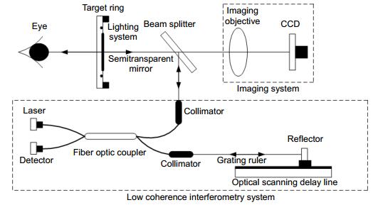
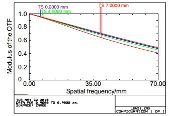
In order to reduce the alignment deviation of the imaging keratometer along the optical axis and improve the measurement accuracy of corneal diopter, a high precision imaging keratometer optical system was designed. The optical system includes imaging system and low coherence interferometry system. The imaging system consists of imaging objective, cornea, and measurement target ring, wherein the imaging objective lens adopts a double telocentric optical path design. The low-coherence interferometry system uses the grating scale to measure the displacement of the scanning mirror, and then locates the vertices of the cornea and the measuring target ring by low-coherence interference signals, achieving accurate measurement between the apex of the cornea and the distance of the measuring target ring. The imaging objective has a modulation transfer function greater than 0.4 at a maximum spatial frequency of 70 lp/mm, the distortion is less than 0.05%. The simulation results show that the system has a compact structure, good imaging quality and simple operation. It meets the demand for high precision measurement of corneal refractive power by an imaging keratometer. -

-
References
[1] Rogers D L, Whitehead G R, Stephens J A, et al. Corneal power measurements in fixating versus anesthetized nonfixating children using a handheld keratometer[J]. Journal of American Association for Pediatric Ophthalmology and Strabismus, 2010, 14(1): 11-14. doi: 10.1016/j.jaapos.2009.11.008 [2] 赵俊奇, 段培华, 郭智勇, 等.人眼角膜曲率参数亚像素测量系统的设计[J].中北大学学报(自然科学版), 2011, 32(3): 362-366. doi: 10.3969/j.issn.1673-3193.2011.03.022 Zhao J Q, Duan P H, Guo Z Y, et al. Design of subpixel algorithm of dioptric paramater measurement system for eye cornea[J]. Journal of North University of China (Natural Science Edition), 2011, 32(3): 362-366. doi: 10.3969/j.issn.1673-3193.2011.03.022 [3] Karnowski K, Kaluzny B J, Szkulmowski M, et al. Corneal topography with high-speed swept source OCT in clinical examination[J]. Biomedical Optics Express, 2011, 2(9): 2709-2720. doi: 10.1364/BOE.2.002709 [4] Howland H C, Howland B. Photorefraction: a technique for study of refractive state at a distance[J]. Journal of the Optical Society of America, 1974, 64(2): 240-249. doi: 10.1364/JOSA.64.000240 [5] 李明东, 高兴宇, 陈朋波, 等.基于机器视觉的高分辨率双远心物镜设计[J].光学仪器, 2016, 38(3): 226-232. doi: 10.3969/j.issn.1005-5630.2016.03.007 Li M D, Gao X Y, Chen P B, et al. The design of high resolution double telecentric lens based on machine vision[J]. Optical Instruments, 2016, 38(3): 226-232. doi: 10.3969/j.issn.1005-5630.2016.03.007 [6] 李明东, 高兴宇, 叶鹏, 等.机器视觉非球面双远心物镜的设计[J].激光与光电子学进展, 2016, 53(7): 70801. Li M D, Gao X Y, Ye P, et al. Design of aspherical double telecentric lens for machine vision[J]. Laser & Optoelectronics Progress, 2016, 53(7): 70801. [7] 夏兵, 王敏, 郭巧双, 等.用于小零件图像测量的双远心光学系统[J].光学仪器, 2015, 37(4): 314-318. doi: 10.3969/j.issn.1005-5630.2015.04.007 Xia B, Wang M, Guo Q S, et al. Bilateral telecentric system for image measurement of small parts[J]. Optical Instruments, 2015, 37(4): 314-318. doi: 10.3969/j.issn.1005-5630.2015.04.007 [8] 刘富国, 查学军, 杨波, 等.基于光纤低相干干涉技术的透镜中心厚度测量方法研究[J].应用激光, 2016, 36(5): 605-610. Liu F G, Zha X J, Yang B, et al. Study on the method of measuring the center thickness of the lenses based on low coherence interferometry of optical fiber[J]. Applied Laser, 2016, 36(5): 605-610. [9] 师中华, 杨宝喜, 胡小邦, 等.基于低相干干涉技术的大量程高精度镜面间距测量[J].光学学报, 2016, 36(6): 612001. Shi Z H, Yang B X, Hu X B, et al. Lens surface distance measurement with large range and high precision based on low coherence interferometry[J]. Acta Optica Sinica, 2016, 36(6): 612001. [10] 郑少林, 刘永基, 王肇圻, 等.新型成像角膜曲率仪的光学系统设计[J].光学学报, 2013, 33(5): 522004. Zheng S L, Liu Y J, Wang Z Q, et al. Design of optical system for a novel imaging keratometer[J]. Acta Optica Sinica, 2013, 33(5): 522004. [11] 隋成华, 沃圣杰, 高楠, 等.基于Placido盘的角膜地形图仪成像系统设计与实现[J].光学学报, 2016, 36(12): 1222001. Sui C H, Wo S J, Gao N, et al. Design and implementation of imaging system for corneal topography based on Placido disk[J]. Acta Optica Sinica, 2016, 36(12): 1222001. [12] 赵俊奇, 郭智勇, 陈安世, 等.一种基于图像处理的人眼全自动角膜曲率计研究[J].中国生物医学工程学报, 2011, 30(1): 100-104. doi: 10.3969/j.issn.0258-8021.2011.01.017 Zhao J Q, Guo Z Y, Chen A S, et al. Auto-ophthalmometer of eye based on image processing[J]. Chinese Journal of Biomedical Engineering, 2011, 30(1): 100-104. doi: 10.3969/j.issn.0258-8021.2011.01.017 [13] 闫洁, 孟鹏花, 赵俊奇.人眼角膜曲率测量系统的研究[J].应用基础与工程科学学报, 2011, 19(S1): 254-261. Yan J, Meng P H, Zhao J Q. Research of curvature measuring system of eyes cornea[J]. Journal of Basic Science and Engineering, 2011, 19(S1): 254-261. -
Overview

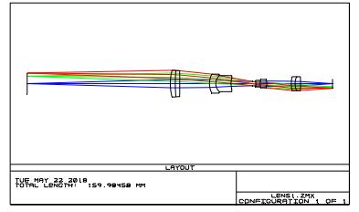
Overview: In order to reduce the alignment deviation of the imaging keratometer along the optical axis and improve the measurement accuracy of corneal diopter, a high precision imaging keratometer optical system was designed. The optical system includes imaging system and low coherence interferometry system. The imaging system consists of imaging objective, cornea, and measurement target ring. Imaging objective lens consists of double telocentric light path, which has large depth of field, low distortion and constant magnification in a certain object distance range. The magnification of the image stays the same with any object distance changes within a certain object distance range. The change of image distance does not affect the size of the image as well. It also has advantages of being insensitive to the object distance and the image distance change contribute to the image obtained by different human eyes and the magnification stability when the human eye moves slightly because of the relatively large depth of field. It is convenient and quick to align in the direction of the optical axis during measurement. The low-coherence interferometry system uses the grating scale to measure the displacement of the scanning mirror, and then locates the vertices of the cornea and the measuring target ring by low-coherence interference signals, achieving accurate measurement between the apex of the cornea and the distance of the measuring target ring. The use of low coherence interferometry solves the problem of using the double telocentric lens in the imaging keratometer which is hardly determining the distance from the apex of the cornea to the measuring target ring accurately, and improves the measurement accuracy of this distance. The design completed system has the following parameters, the total length of the imaging objective system is 85 mm, the object height is 14 mm, the image height is 6.2 mm, the magnification is -0.4, the depth of field is 8 mm, the spectral range is 900 nm~980 nm, and the maximum spatial frequency is 70 lp/mm. The modulation transfer function at mm is greater than 0.4, and the distortion is less than 0.05%. The parameters meet the design requirements of corneal index. The low-coherence interferometry system uses a broadband light source with a center wavelength of 850 nm, a 2×2 fiber coupler with a split ratio of 99:1, and a linear delay line with a scan speed of 120 mm/s to achieve precise positioning of the cornea. The system has compact structure, good imaging quality and simple operation, and meets the high-precision measurement requirements of imaging keratometer for corneal diopter. In summary, the system has practical significance for realizing high-precision corneal curvature measurement.
-
Access History
Figures(10)
Tables(5)
Article Metrics


Export File
Citation
Zhang Xueying, Wang Jinsong, Huang Guolin, et al. Design of optical system for high accuracy imaging keratometry[J]. Opto-Electronic Engineering, 2019, 46(1): 180392. doi: 10.12086/oee.2019.180392
Format
Content
 DownLoad:


-
Figure 1.
High-precision imaging keratometer optical system schematic
-
Figure 2.
Double telecentric lens imaging schematic
-
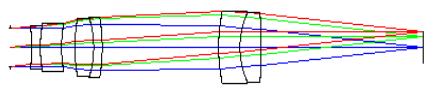
Figure 3.
Initial structure diagram
-
Figure 4.
Double telecentric imaging objective lens structure diagram
-
Figure 5.
MTF graph
-
Figure 6.
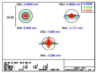
Plot diagram
-
Figure 7.
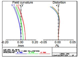
Field curvature and distortion
-
Figure 8.
PSF graph
-
Figure 9.
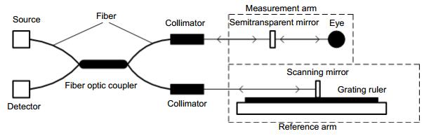
Low coherence interferometry system
-
Figure 10.
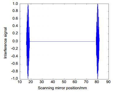
Interference signal simulation diagram


