
 E-mail Alert
 RSS
RSS
-
摘要:
自适应光学技术能够对波前像差进行实时测量及调控。1997年,该技术被首次成功地应用于活体人眼像差的调控,并获得了接近衍射极限的高分辨力视网膜视细胞图像和传统低阶像差矫正无法达到的“超视力”。随后自适应光学技术在眼科学研究中得到迅速发展。就研究内容来看,该领域主要包括视网膜高分辨力成像和人眼像差操控与视功能研究两大方向。美国Rochester大学Williams教授和加利福利亚大学Roorda教授于2011年分别对视网膜高分辨力成像和人眼像差操控与视功能研究方向的研究作了非常全面的综述。1997年,光电所在国内率先开展人眼自适应光学技术及其应用研究,本文在简单介绍人眼自适应光学系统原理的基础上,报道了光电所在该领域近五年的主要研究进展。
Abstract:Adaptive optics (AO) techniques allow to measure and manipulate the wavefront aberrations in real time. This technology was first successfully applied to the manipulation of ocular aberrations, and high resolution retinal images and "supernormal vision" were achieved in vivo for the first time in 1997. Subsequently, adaptive optics were developed rapidly in ophthalmology. This field includes two main directions: high resolution imaging of the retina and the research of ocular aberrations manipulation and its influence on visual function. In 2011, professor D. R. Williams from Rochester University and Professor Austin Roorda from California University made two comprehensive overviews on retinal high-resolution imaging and ocular aberrations manipulation and its influence on visual function, respectively. In 1997, reseachers in Institute of Optics and Electronics took the lead in developing of adaptive optics technology in China. This article firstly introduces the principle of ocular adaptive optical system briefly, and then reports the main research progress of Institute of Optics and Electronics in this field for the last five years.
-
Key words:
- ocular aberrations /
- adaptive optics /
- visual training /
- vision simulation /
- intraocular scatter /
- image restoration
-

Overview: Adaptive optics (AO) techniques allow to measure and manipulate the wavefront aberrations in real time. In 1997, reseachers in Institute of Optics and Electronics (IOE) took the lead in developing of adaptive optics technology in China. This article firstly introduces the principle of ocular adaptive optical system briefly, and then reports the main research progress of IOE for the last five years. In 2012, we measured the effects of perceptual learning on visual sensitivity with and without HOAs-correction both for normal adults, and older child and adult anisometropic amblyopes. We found larger and more robust contrast sensitivity improvements when the HOAs were corrected than when they were left uncorrected. In 2014, we investigated the effects of ocular aberrations on the binocular vision functions. Results showed that ocular aberrations had an inequable effect on the different binocular vision functions. In 2016, an adaptive optics double pass (DP) system was designed and developed for ocular scatter estimation. The experimental results showed that this system could improve the accuracy of ocular scatter estimation. Then we investigated the effect of HOAs and intraocular scatter on contrast sensitivity. Quantitative analysis suggested a potential compensatory mechanism between HOAs and intraocular scatter on contrast sensitivity. In 2016, an AO pattern reversal visual evoked potential (PR-VEP) measurement system was established. PR-VEP measurements were performed with HOAs either retained or corrected. The results confirmed the contributions of the HOAs to the alterations in PR-VEP, and suggested that HOAs should be corrected to realize accurate PR-VEP testing. In 2017, we proposed a deep learning method to restore the degraded retinal images. The method directly learned an end-to-end mapping between the blurred and restored retinal images. The mapping was represented as a deep convolutional neural network. This network was validated on synthetically generated retinal images as well as real AO retinal images. The assessment of the restored retinal images demonstrated that the image quality had been significantly improved.
-

-
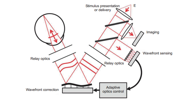
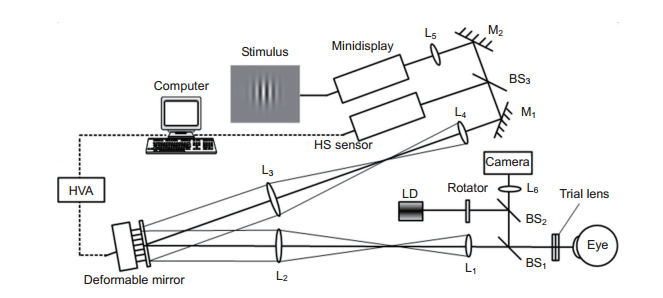
图 2 人眼像差自适应光学矫正与视知觉学习训练系统原理图
Figure 2. Schematic diagram of adaptive optical perceptual learning system
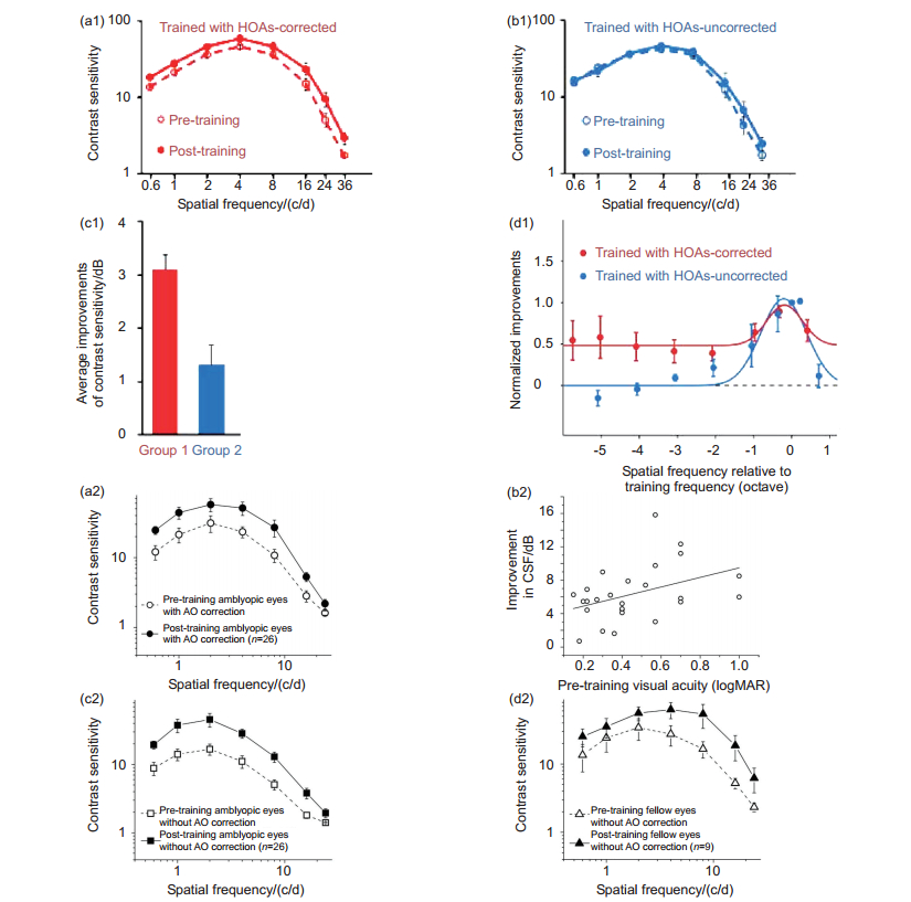
图 3 (a1)~(d1)正常成年人眼在高阶像差矫正与不矫正下进行知觉学习训练后的对比敏感度及其收益[8]。(a1)高阶像差校正下的学习训练结果;(b1)非高阶像差校正下的学习训练结果;(c1)两组训练后平均对比敏感度提高的幅度;(d1)两组训练后不同空间频率下的归一化改善程度。(a2)~(d2)弱视患者人眼在高阶像差矫正与不矫正下进行知觉学习训练后的对比敏感度及其收益[9]。(a2)高阶像差校正下的学习训练结果;(b2)高阶像差校正训练后对比敏感度收益与训练前视锐度的函数关系; (c2)非高阶像差校正下的学习训练结果;(d2)非高阶像差校正下的学习训练后另一只眼睛的对比敏感度测试结果
Figure 3. (a1)~(d1) Contrast sensitivity function and its benefit for normal adults pre-and post-training with and without correction of high-order aberrations (HOAs). (a1) Average results trained under the HOAs-corrected condition[8]; (b1) Average post-and pre-training CSFs without HOAs-corrected; (c1) Average improvements of contrast sensitivity with and without HOAs-corrected; (d1) Average magnitude of contrast sensitivity improvements across observers and spatial frequencies. (a2)~(d2) Improvements in the CSF after perceptual learning for older children and adults with anisometropic amblyopia. (a2) Pre-and post-training CSFs for amblyopic eyes with AO correction[9]; (b2) Average improvements in CSF with AO correction (dB) as a function of pre-training amblyopic VA (logMAR); (c2) The learning effect with AO correction is maintained under the condition without AO correction for the amblyopic eyes; (d2) The learning effect with AO correction is transferred to the untrained fellow eyes

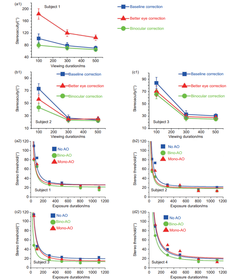
图 5 (a1)~(c1)不同视标显示时间和像差校正策略下的立体视阈值[11]。图中Baseline correction代表仅校正低价像差的结果;Better eye corection代表优势眼像差校正的结果;Binocular correction代表双眼高阶像差校正的结果。(a2)~(d2)二次叠加模型拟合后的立体视阈值-显示时间曲线[12]。图中No AO代表仅校正低价像差的结果;Bino-AO代表双眼高阶像差校正的结果;Mono-AO代表优势眼像差校正的结果
Figure 5. (a1)~(c1) Stereoacuity versus viewing duration under different optical conditions for the three subjects[11]. Blue squares represent stereoacuity with baseline correction, green circles and red triangles represent stereoacuity with binocular and better eye correction. (a2)~(d2) Stereo threshold versus exposure duration under different optical conditions for the 4 subjects on linear scales[12]. The blue squares represent stereoacuity with basic correction of defocus and astigmatism, the green circles and red triangles represent stereoacuity with binocular and better eye correction

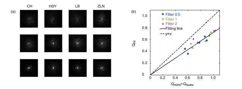
图 7 (a) 四名被试者在三种像差校正策略下的双通PSF测量典型结果[22],每列对应一个被试者的结果,其中第一行是仅校正低价像差获得结果,第二行是校正前五阶像差后获得的结果,第三行是校正前八阶像差后获得的结果;(b)像差和散射同时存在时的影响因子(Q)与两者单独存在时的影响因子(Q)之和的线性拟合结果[23],其中三种不同颜色对应的结果分别是采用三种散射滤波片获得的实验结果,拟合结果为y=0.73x。
Figure 7. (a) The typical DP PSF images measured at the fovea for the left eyes of subjects in AO-DPPMS system with different aberration correction strategies[23]. First row, DP images with defocus and astigmatism corrected in the AO-DPPMS system; second row, DP images with aberrations corrected up to the 5th Zernike order with AO-DPPMS system; third row, DP images with aberrations corrected up to the 8th Zernike order with AO-DPPMS system; (b) Linear fit of impact factorHS (QHS) versus the sum of impact factorHOAs (QHOAS) and impact factorScatter (QScatter). Each point represent an average value across subjects for a spatial frequency. Blue circles stand for results obtained with filter S0.5; green triangles stand for results obtained with filter S1; magenta diamonds stand for results obtained with filter S2. The solid line represent the fitting line; the dotted line stand for the line y = x
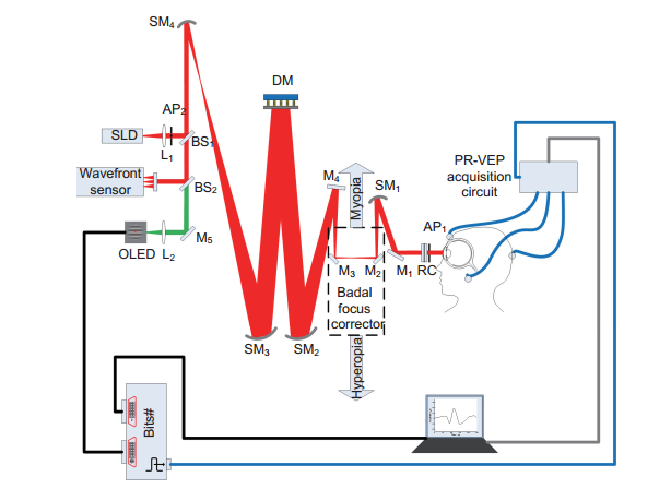
图 8 自适应光学客观视力检查系统原理图
Figure 8. AO system integrated visual performance evaluation with pattern reversal VEP measurements
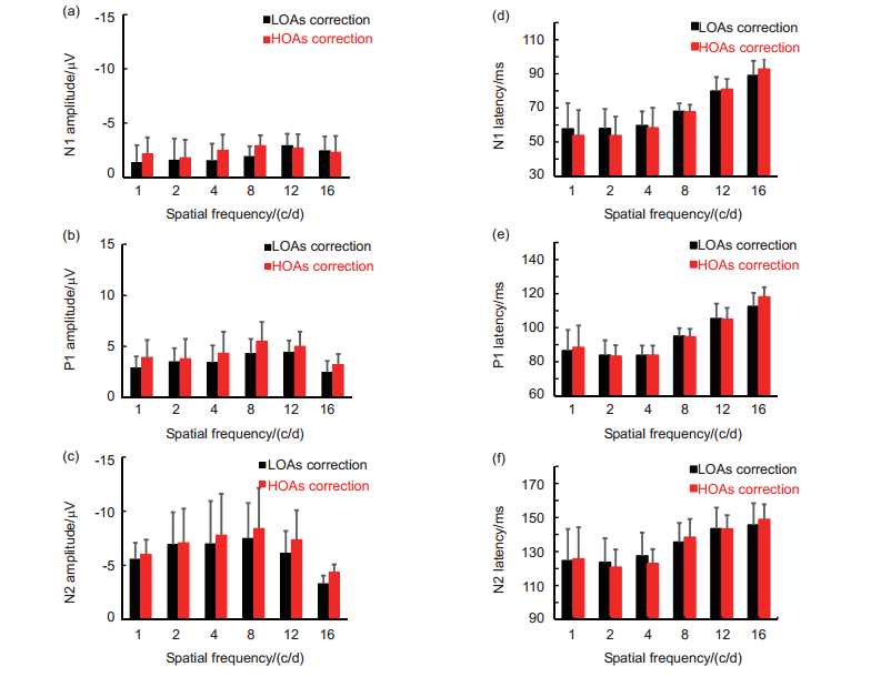
图 9 低阶像差和高阶像差矫正下VEP检查的平均结果
Figure 9. Average VEP magnitude with low-order aberrations corrected and with high-order aberrations corrected
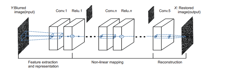
图 10 网络结构示意图。降质图像经过网络特征提取、非线性映射及重构后获得复原图像,网络中的参数通过学习获得
Figure 10. Network structure. A blurred image goes through layers and transforms into a restored one, we use 64 filters for each convolutional layer and some sample feature maps are drawn for visualization
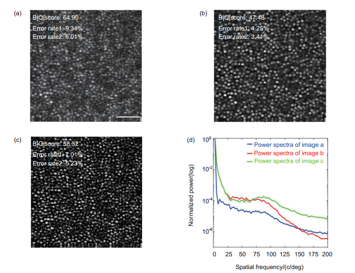
图 11 视网膜图像处理结果。(a)原始图像;(b)使用神经网络处理结果;(c)使用传统盲目反卷积算法处理结果;(d)相应图像归一化功率谱,尺度为50 µm
Figure 11. Deconvolution results of a retinal image captured by AOSLO system. (a) Original image (0.9 mm eccentricity from the foveal center); (b) Restored by the proposed method; (c) Restored by the ALM method; (d) The corresponding normalized image power spectra, the scale bar is 50 µm
-
[1] Liang J Z, Williams D R. Aberrations and retinal image quality of the normal human eye[J]. Journal of the Optical Society of America A, 1997, 14(11): 2873-2883. doi: 10.1364/JOSAA.14.002873
[2] Williams D R. Imaging single cells in the living retina[J]. Vision Research, 2011, 51(13): 1379-1396, doi: 10.1016/j.visres.2011. 05.002.
[3] Roorda A. Adaptive optics for studying visual function: A comprehensive review[J]. Journal of Vision, 2011, 11(5): 6. doi: 10.1167/11.5.6
[4] Rossi E A, Weiser P, Tarrant J, et al. Visual performance in emmetropia and low myopia after correction of high-order aberrations[J]. Journal of Vision, 2007, 7(8): 14. doi: 10.1167/7.8.14
[5] Chen L, Artal P, Gutierrez D, et al. Neural compensation for the best aberration correction[J]. Journal of Vision, 2007, 7(10): 9. doi: 10.1167/7.10.9
[6] Elliott S L, Choi S S, Doble N, et al. Role of high-order aberrations in senescent changes in spatial vision[J]. Journal of Vision, 2009, 9(2): 24. doi: 10.1167/9.2.24
[7] Dai Y, Zhao L N, Xiao F, et al. Adaptive optics vision simulation and perceptual learning system based on a 35-element bimorph deformable mirror[J]. Applied Optics, 2015, 54(5): 979-985. doi: 10.1364/AO.54.000979
[8] Zhou J W, Zhang Y D, Dai Y, et al. The eye limits the brain's learning potential[J]. Scientific Reports, 2012, 2: 364, doi: 10.1038/srep00364.
[9] Liao M, Zhao H X, Liu L Q, et al. Training to improve contrast sensitivity in amblyopia: correction of high-order aberrations[J]. Scientific Reports, 2016, 6: 35702, doi: 10.1038/srep35702.
[10] 梁波. 自适应光学像差矫正对双眼叠加作用的影响研究[D]. 成都: 中国科学院光电技术研究所, 2013.
Liang B. Effects of adaptive optics correction of ocular aberrations on binocular summation[D]. Chengdu: Institute of Optics and Electronics, Chinese Academy of Sciences, 2013.
http://www.wanfangdata.com.cn/details/detail.do?_type=degree&id=Y2368223 [11] Kang J, Xiao F, Zhao J L, et al. Effects of higher-order aberration correction on stereopsis at different viewing durations[J]. Journal of Biomedical Optics, 2015, 20(7): 075005. doi: 10.1117/1.JBO.20.7.075005
[12] Kang J, Dai Y, Zhang Y D. Temporal integration property of stereopsis after higher-order aberration correction[J]. Biomedical Optics Express, 2015, 6(11): 4472-4482. doi: 10.1364/BOE.6.004472
[13] 康健. 人眼高阶像差校正对立体视觉和双眼调节的影响研究[D]. 成都: 中国科学院光电技术研究所, 2016.
Kang J. Effect of ocular higher-order aberration correction on stereopsis and binocular accommodation[D]. Chengdu: Institute of Optics and Electronics, Chinese Academy of Sciences, 2016.
http://cdmd.cnki.com.cn/Article/CDMD-80151-1016757885.htm [14] IJspeert J K, de Waard P W T, van den Berg T J T P, et al. The intraocular straylight function in 129 healthy volunteers; Dependence on angle, age and pigmentation[J]. Vision Research, 1990, 30(5): 699-707. doi: 10.1016/0042-6989(90)90096-4
[15] Artal P, Benito A, Pérez G M, et al. An objective scatter index based on double-pass retinal images of a point source to classify cataracts[J]. PLoS One, 2011, 6(2): e16823. doi: 10.1371/journal.pone.0016823
[16] Güell J L, Pujol J, Arjona M, et al. Optical quality analysis system; instrument for objective clinical evaluation of ocular optical quality[J]. Journal of Cataract and Refractive Surgery, 2004, 30(7): 1598-1599. doi: 10.1016/j.jcrs.2004.04.031
[17] Benito A, Pérez G M, Mirabet S, et al. Objective optical assessment of tear-film quality dynamics in normal and mildly symptomatic dry eyes[J]. Journal of Cataract & Refractive Surgery, 2011, 37(8): 1481-1487. http://www.sciencedirect.com/science/article/pii/S0886335011007322
[18] Nanavaty M A, Stanford M R, Sharma R, et al. Use of the double-pass technique to quantify ocular scatter in patients with uveitis: A pilot study[J]. Ophthalmologica, 2011, 225(1): 61-66. doi: 10.1159/000316690
[19] Piñero D P, Ortiz D, Alio J L. Ocular scattering[J]. Optometry and Vision Science: Official Publication of the American Academy of Optometry, 2010, 87(9): E682-E96. doi: 10.1097/OPX.0b013e3181e87da6
[20] Artal P, Iglesias I, López-Gil N, et al. Double-pass measurements of the retinal-image quality with unequal entrance and exit pupil sizes and the reversibility of the eye's optical system[J]. Journal of the Optical Society of America A, 1995, 12(10): 2358-2366. doi: 10.1364/JOSAA.12.002358
[21] 赵军磊. 人眼散射客观评价及其对视功能影响研究[D]. 成都: 中国科学院光电技术研究所, 2017.
Zhao J L. Objective assessment of ocular scatter and its influence on visual function[D]. Chengdu: Institute of Optics and Electronics, Chinese Academy of Sciences, 2017.
http://ir.ioe.ac.cn/handle/181551/8166?null [22] Zhao J L, Xiao F, Kang J, et al. Quantifying intraocular scatter with near diffraction-limited double-pass point spread function[J]. Biomedical Optics Express, 2016, 7(11): 4595-4604. doi: 10.1364/BOE.7.004595
[23] Zhao J L, Xiao F, Zhao H X, et al. Effect of higher-order aberrations and intraocular scatter on contrast sensitivity measured with a single instrument[J]. Biomedical Optics Express, 2017, 8(4): 2138-2147. doi: 10.1364/BOE.8.002138
[24] Xiao F, Zhao J L, Zhao H X, et al. Deblurring adaptive optics retinal images using deep convolutional neural networks[J]. Biomedical Optics Express, 2017, 8(12): 5675-5687. doi: 10.1364/BOE.8.005675
-
 下载:
下载:
 点击扫一扫
点击扫一扫
图(11)
计量
- 文章访问数: 12358
- PDF下载数: 4254
- 施引文献: 0


