
 E-mail Alert
 RSS
RSS
| Citation: |
Chen L W, Zhou Y, Wu M X, Hong M H. Remote-mode microsphere nano-imaging: new boundaries for optical microscopes. Opto-Electron Adv 1, 170001 (2018). doi: 10.29026/oea.2018.170001

|
Remote-mode microsphere nano-imaging: new boundaries for optical microscopes
-
Abstract
Optical microscope is one of the most popular characterization techniques for general purposes in many fields. It is distinguished from the vacuum or tip-based imaging techniques for its flexibility, low cost, and fast speed. However, its resolution limits the functionality of current optical imaging performance. While microspheres have been demonstrated for improving the observation power of optical microscope, they are directly deposited on the sample surface and thus the applications are greatly limited. We develop a remote-mode microsphere nano-imaging platform which can scan freely and in real-time across the sample surfaces. It greatly increases the observation power and successfully characterizes various practical samples with the smallest feature size down to 23 nm. This method offers many unique advantages, such as enabling the detection to be non-invasive, dynamic, real-time, and label-free, as well as leading to more functionalities in ambient air and liquid environments, which extends the nano-scale observation power to a broad scope in our life.-
Keywords:
- nano-imaging /
- label-free /
- microscopy
-

-
References
[1] Clevelanda J, Montvillea T J, Nesb I F, Chikindas M L. Bacteriocins: safe, natural antimicrobials for food preservation. Int J Food Microbiol 71, 1–20 (2001). doi: 10.1016/S0168-1605(01)00560-8 [2] O'Mara W C, Herring R B, Hunt L P. Handbook of Semiconductor Silicon Technology (Noyes Publications, 1990). [3] Bradbury S. The microscope: past and present (Pergamon Press, 1968). [4] Qin F, Huang K, Wu J F, Teng J H, Qiu C W et al. A supercritical lens optical label-free microscopy: sub-diffraction resolution and ultra-long working distance. Adv Mater 29, 1602721 (2017). doi: 10.1002/adma.201602721 [5] Qin F, Ding L, Zhang L, Monticone F, Chum C C et al. Hybrid bilayer plasmonic metasurface efficiently manipulates visible light. Sci Adv 2, 1–9 (2016). [6] Qin F, Huang K, Wu J F, Jiao J, Luo X G et al. Shaping a subwavelength needle with ultra-long focal length by focusing azimuthally polarized light. Sci Rep 5, 9977 (2015). doi: 10.1038/srep09977 [7] Wang Y T, Cheng B H, Ho Y Z, Lan Y C, Luan P G et al. Gain-assisted hybrid-superlens hyperlens for nano imaging. Opt Express 20, 22953–22960 (2012). doi: 10.1364/OE.20.022953 [8] Lin Y H, Tsai D P. Near-field scanning optical microscopy using a super-resolution cover glass slip. Opt Express 20, 16205–16211 (2012). doi: 10.1364/OE.20.016205 [9] Fukaya T, Buchel D, Shinbori S, Tominaga J, Atoda N et al. Micro-optical nonlinearity of a silver oxide layer. J Appl Phys 89, 6139–6144 (2001). doi: 10.1063/1.1365434 [10] Li Y, Li X, Chen L W, Pu M B, Jin J J et al. Orbital angular momentum multiplexing and demultiplexing by a single metasurface. Adv Opt Mater 5, 1600502 (2017). [11] Li X, Chen L W, Li Yang, Zhang X H, Pu M B et al. Multicolor 3D meta-holography by broadband plasmonic modulation. Sci Adv 2, e1601102 (2016). doi: 10.1126/sciadv.1601102 [12] Huang B, Wang W Q, Bates M, Zhuang X W. Three-dimensional super-resolution imaging by stochastic optical reconstruction microscopy. Science 319, 810–813 (2008). doi: 10.1126/science.1153529 [13] Betzig E, Patterson G H, Sougrat R, Lindwasser O W, Olenych S et al. Imaging intracellular fluorescent proteins at nanometer resolution. Science 313, 1642–1645 (2006). doi: 10.1126/science.1127344 [14] Binnig G, Rohrer H. Scanning tunneling microscopy. IBM J Res Dev 4, 355–369 (1986). [15] Luo X. Principles of electromagnetic waves in metasurfaces. Sci China: Phys Mech Astron 58, 594201 (2015). doi: 10.1007/s11433-015-5688-1 [16] Wang Y T, Cheng B H, Ho Y Z, Lan Y C, Luan P G et al. Optical hybrid-superlens hyperlens for superresolution imaging. IEEE J Sel Top Quantum Electron 19, 4601305 (2013). doi: 10.1109/JSTQE.2012.2230152 [17] Cheng B H, Lan Y C, Tsai D P. Breaking optical diffraction limitation using optical hybrid-super-hyperlens with radially polarized light. Opt Express 21, 14898–14906 (2013). doi: 10.1364/OE.21.014898 [18] Luo X, Ishihara T. Surface plasmon resonant interference nanolithography technique. Appl Phys Lett 84, 4780–4782 (2004). doi: 10.1063/1.1760221 [19] Tang D, Wang C, Zhao Z, Wang Y, Pu M et al. Ultrabroadband superoscillatory lens composed by plasmonic metasurfaces for subdiffraction light focusing. Laser Photonics Rev 9, 713–719 (2015). doi: 10.1002/lpor.201500182 [20] Editorial. So Much More to Know. Science 309, 78–102 (2005). [21] Yu N, Genevet P, Kats M A, Aieta F, Tetienne J -P et al. Light propagation with phase discontinuities: generalized laws of reflection and refraction. Science 334, 333–337 (2011). doi: 10.1126/science.1210713 [22] Pu M, Li X, Ma X, Wang Y, Zhao Z et al. Catenary optics for achromatic generation of perfect optical angular momentum. Sci Adv 1, e1500396 (2015). doi: 10.1126/sciadv.1500396 [23] Li X, Pu M, Wang Y, Ma X, Li Y et al. Dynamic control of the extraordinary optical scattering in semicontinuous 2d metamaterials. Adv Opt Mater 4, 659–663 (2016). [24] Wang Z B, Guo W, Li L, Luk'yanchuk B, Khan A, Liu Z et al. Optical virtual imaging at 50 nm lateral resolution with a white-light nanoscope. Nat Commun 2, 218 (2011). doi: 10.1038/ncomms1211 [25] Yang H, Trouillon R, Huszka G, Gijs M A M. Super-resolution imaging of a dielectric microsphere is governed by the waist of its photonic nanojet. Nano Lett 16, 4862–4870 (2016). doi: 10.1021/acs.nanolett.6b01255 [26] Darafsheh A, Guardiola C, Palovcak A, Finlay J C, Cárabe A. Optical super-resolution imaging by high-index microspheres embedded in elastomers. Opt Lett 40, 5–8 (2015). doi: 10.1364/OL.40.000005 [27] Wang F F, Liu L Q, Yu P, Liu Z, Yu H B et al. Three-dimensional super-resolution morphology by near-field assisted white-Light interferometry. Sci Rep 6, 24703 (2016). doi: 10.1038/srep24703 [28] Darafsheh A, Limberopoulos N I, Derov J S, Walker D E, Astratov V N. Advantages of microsphere-assisted super-resolution imaging technique over solid immersion lens and confocal microscopies. Appl Phys Lett 104, 61117 (2014). doi: 10.1063/1.4864760 [29] Wang F F, Liu L Q, Yu H B, Wen Y D, Yu P et al. Scanning superlens microscopy for non-invasive large field-of-view visible light nanoscale imaging. Nat Commun 7, 13748 (2016). doi: 10.1038/ncomms13748 [30] Born M, Wolf E. Principles of optics; electromagnetic theory of propagation, interference, and diffraction of light (Pergamon Press, 1975). [31] Soroko L M. Meso-optics, foundations and applications (World Scientific, 1996). [32] Hong M, Wu M. Immersion Nanoscope Lens Assembly Chip for Super-resolution Imaging. PRV 10201608343Y (2016). [33] Dunny G M, Brickman T J, Dworkin M. Multicellular behavior in bacteria, communication, cooperation, competition and cheating. Bio Essays 30, 296–298 (2008). -
Supplementary Information
-
Access History

Figures(3)
Article Metrics


Export File
Citation
Chen L W, Zhou Y, Wu M X, Hong M H. Remote-mode microsphere nano-imaging: new boundaries for optical microscopes. Opto-Electron Adv 1, 170001 (2018). doi: 10.29026/oea.2018.170001
Format
Content
 DownLoad:
-
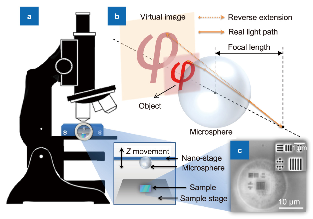
Figure 1.
(a) Schematic diagram of the remote mode optical microsphere setup. (b) Mechanism to illustrate the enlarged virtual image by the microsphere. (c) Optical image captured by this system (Sample: semiconductor testing sample; scale bar: 10 μm; imaged by a 20 μm silica microsphere compiled to an oil-immersion optical microscope with a 100× objective lens, NA=1.4). Inset: SEM image (scale bar: 1 μm).
-
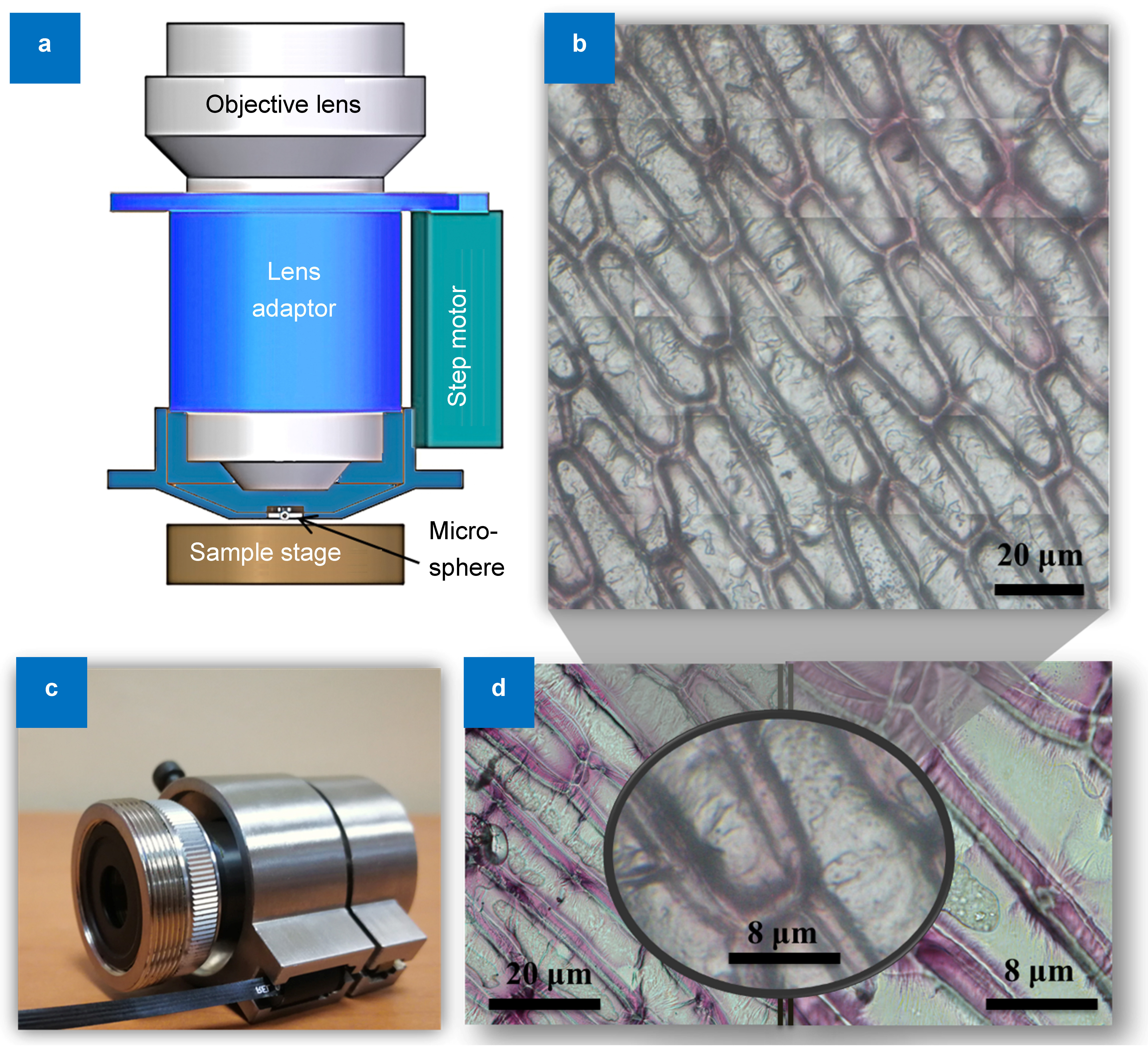
Figure 2.
(a) Schematic of the design of the universal lens adaptor for the microsphere (silica microsphere with 400 μm diameter attached on a 20× objective lens. Characterization was done in ambient air and distance between the onion cell and the microsphere is ~65 μm, with white light illumination). (b) Integrated image of onion cells (scale bar: 20 μm). (c) Optical image of the universal sample adaptor after integration. (d) Comparison of the optical images by three optical lenses: the 20× objective lens (left, scale bar: 20 μm); 20× objective lens with the microsphere (middle, which is our nanoscope design, scale bar: 8 μm); and 50× objective lens (right, scale bar: 8 μm).
-
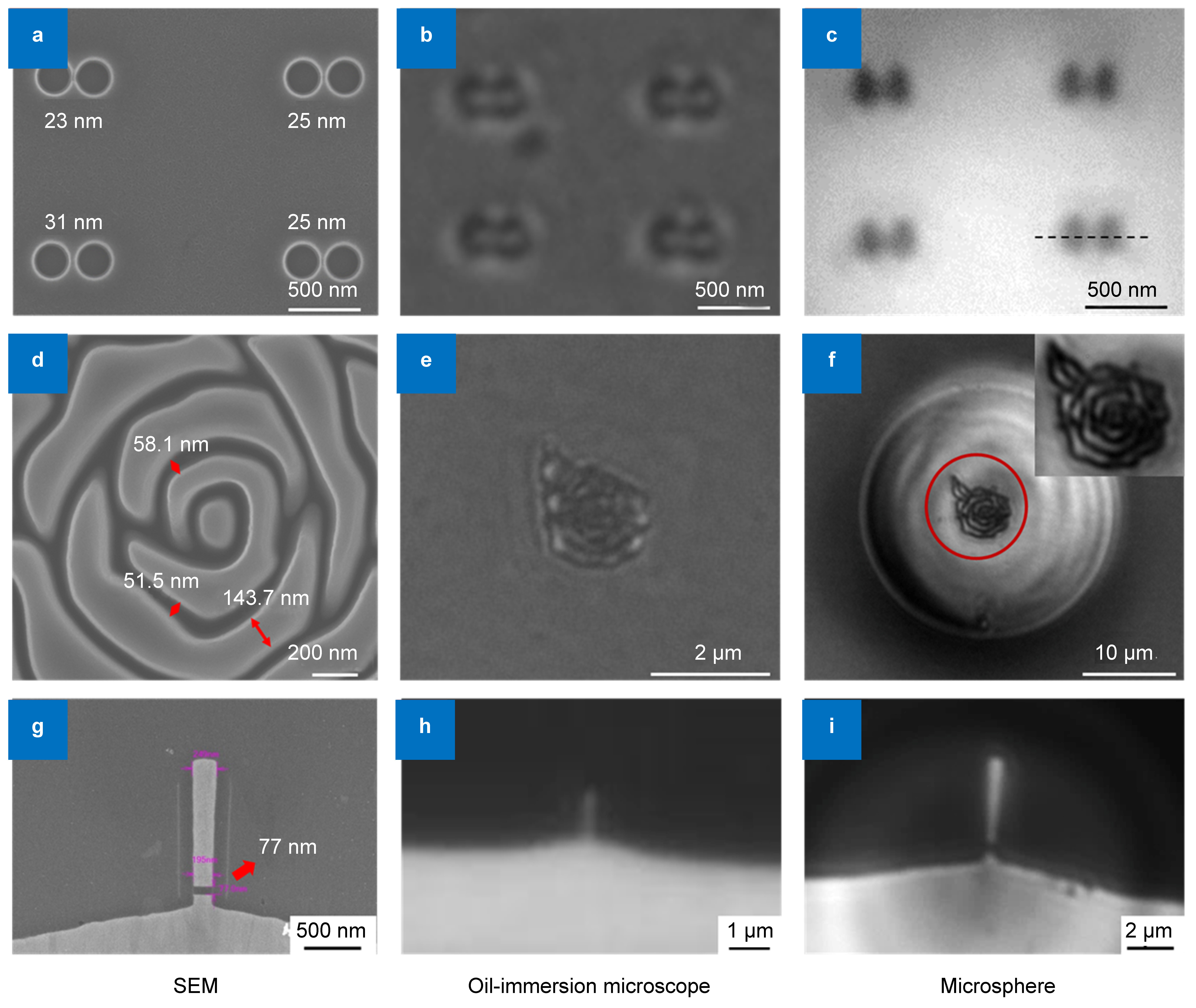
Figure 3.
(a~c) Imaging of nano-dot pairs with nano-gap on a Si wafer. (a) SEM image of the samples, showing sizes of nano-gaps in between each pair of nano-dots. (b) Imaging of the samples by an oil-immersion microscope (neighboring nano-dots cannot be resolved clearly). (c) Neighboring separated nano-dots are resolved clearly by a microsphere with 20 μm diameter. The back dash line in (c) indicates the line cut (the intensity analysis is presented in supplementary materials). (d~f) Imaging of samples with complex features (the "nano-rose"). (d) Zoomed-in SEM image with size notations, it shows that the typical line width of the structure is ~140 nm, and separated by nano-grooves with a typical size ranging from 50~60 nm. (e) Imaging result by the oil-immersion optical microscope. (f) Image under the 27μm microsphere in scanning mode. The diameter of the microsphere is larger in order to contain the entire nano-rose in the central region. (Compared to the microsphere used for the imaging of nano-dots, the microsphere with a larger diameter is chosen to ensure the entire nano-rose pattern is in the central region of the microsphere. Inset: zoomed-in image under the microsphere). (g~i) Imaging of a magnetic head in a hard disc drive from the production line. (g) SEM image of the magnetic head, with a nano-gap of 77 nm. (h) Imaging by a conventional oil-immersion microscope. (i) Imaging by the microsphere nanoscope in non-contact mode. The three columns represent images obtained by SEM, oil-immersion optical microscope (100×, NA 1.4), and microsphere nanoscope, respectively.


